
Pectus Carinatum
The most second common chest wall deformity, pectus carinatum or pigeon chest like pectus excavatum is most likely caused by abnormal growth of the costal cartilage between the ribs and sternum. It usually becomes apparent during the growth spurt and puberty (ages 11-15 years) often seeming to suddenly appear during this period.
Please click on the buttons below to see and read about patients verified experiences (in their own words) and testimonials (which generally include before and after treatment photos). The pectus clinic is very grateful to all the patients who provided feedback.
Symptoms
Similar to pectus excavatum, atypical chest pain and breathlessness on exertion or exercise is often described though of course many patients have no specific symptoms. Chest pain and tenderness are often noted when lying in the prone position (face down). Interestingly, 'asthma' like symptoms are described in up nearly a quarter of teenagers with pectus carinatum. As with pectus excavatum, the effect of patient's self-esteem, body image and confidence can be variable and lead to significant deterioration in mental health.
Video of a CT 3D reconstruction in a young woman with a superior (upper) right pectus carinatum deformity with sternal rotation
Signs
There is little evidence of an obvious association with symptoms of pectus carinatum and signs of a reduction of lung or heart function, though few studies have been conducted. However, patients with pectus carinatum tend to have a 'barrel chested' shape which may alter the mechanics of normal chest wall movement and may explain why some patients experience breathlessness on exertion, further research is needed.
Unlike pectus excavatum, the position of the heart in pectus carinatum is unaffected, however there is an association with a higher incidence of mitral valve prolapse (leaky heart valve) in patients with pigeon chest.
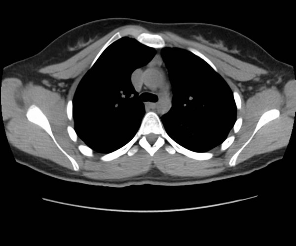
Chest CT scan of a young woman with a less common right asymmetric superior pectus carinatum
Diagnosis
The diagnosis of pectus carinatum is what doctors call a clinical one, that is careful physical examination with a doctor familiar with pectus deformities is usually all that is required.
One of the clinical measurements used to help assess the shape and severity of the chest wall in pectus carinatum has been developed by the Pectus Clinic and isillustrated (to the right). Known as the Cross-Sectional Chest wall ratio or Pectus Index (PI), It allows for an objective measurement to be taken at assessment and during bracing treatment, or before and after surgery. This is demonstrated in the example shown of a 15-year-old boy before and 6 weeks after starting his bracing treatment for his severe pectus carinatum. The nearer the ratio is to 1.0 the 'rounder' the chest (often described as 'barrel chested') the more severe the pectus carinatum deformity.
As with pectus excavatum there is no specific blood test, but radiological assessment (x-rays) may help assess the severity of the pectus carinatum and identify other associated problems such as scoliosis of the spine. The most useful radiological test is a Chest CT scan which allows a more sensitive assessment of the pectus carinatum particularly its severity and should be considered if surgical correction is considered. MRI is an excellent alternative investigation that avoids radiation.
Video of a young man with a severe symmetrical pectus carinatum deformity
Cross sectional Chest Wall Ratio (Pectus Index (PI=ml/ap)
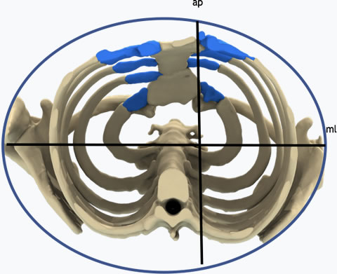
Chest dimensions in a patient with a pectus carinatum deformity
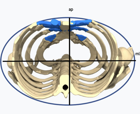
Chest dimensions in a patient without a pectus deformity or following correction
Pectus Index (PI) is where ml is the width of the chest wall (medio-lateral) in centimetres at the level of the nipples and ap is the depth of the chest wall (antero-posterior) between the spine and anterior chest wall at the level of the pectus peak (normal PI ratio 1.5-2.0).
Cross sectional Chest Wall Ratio (Pectus Index (PI=ml/ap)
Chest dimensions in a patient with a pectus carinatum deformity BEFORE BRACING
Chest dimensions in a patient with a pectus carinatum deformity 6 WEEKS AFTER STARTING BRACING TREATMENT
Severity
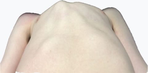
Pectus carinatum usually involves the lower sternal costal cartilages, pushing the sternum forwards and can be symmetrical (bilateral) or often asymmetrical (unilateral) with the right for some reason being more obviously affected. There is usually a compensatory flattening or depression of the rib cage on the opposite side to the pectus if asymmetrical or both sides if symmetrical (hence the alternative term of Keel chest). A rarer from of upper sternal protrusion is also described. There are various types of classification developed to describe the different sorts of pectus carinatum but severity is often based on the extent of sternal protrusion and degree of sternal rotation as well as the extent of compensatory rib flattening. As with pectus excavatum, poor posture often accompanies patients with pectus carinatum as well spinal problems.
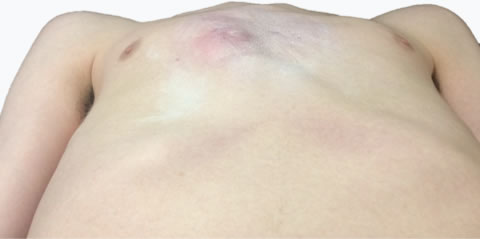
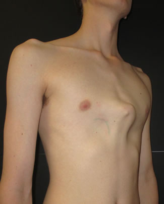
A young boy with a mild asymmetric right pectus carinatum involving lower anterior chest
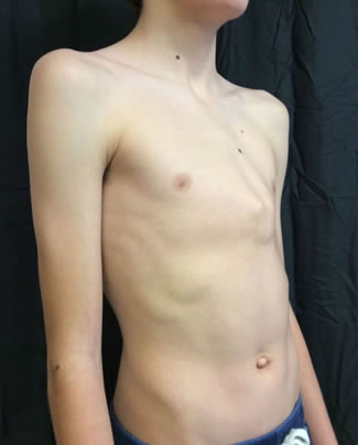
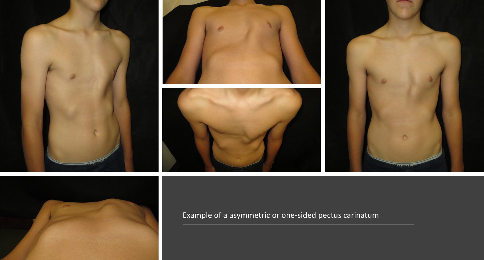
Moderate asymmetric right sided pectus carinatum deformity with mild contralateral left-sided rib flaring
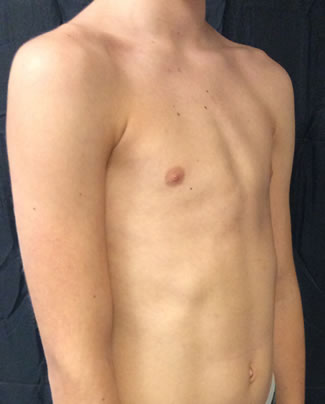
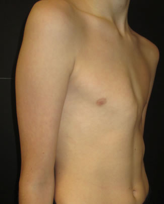
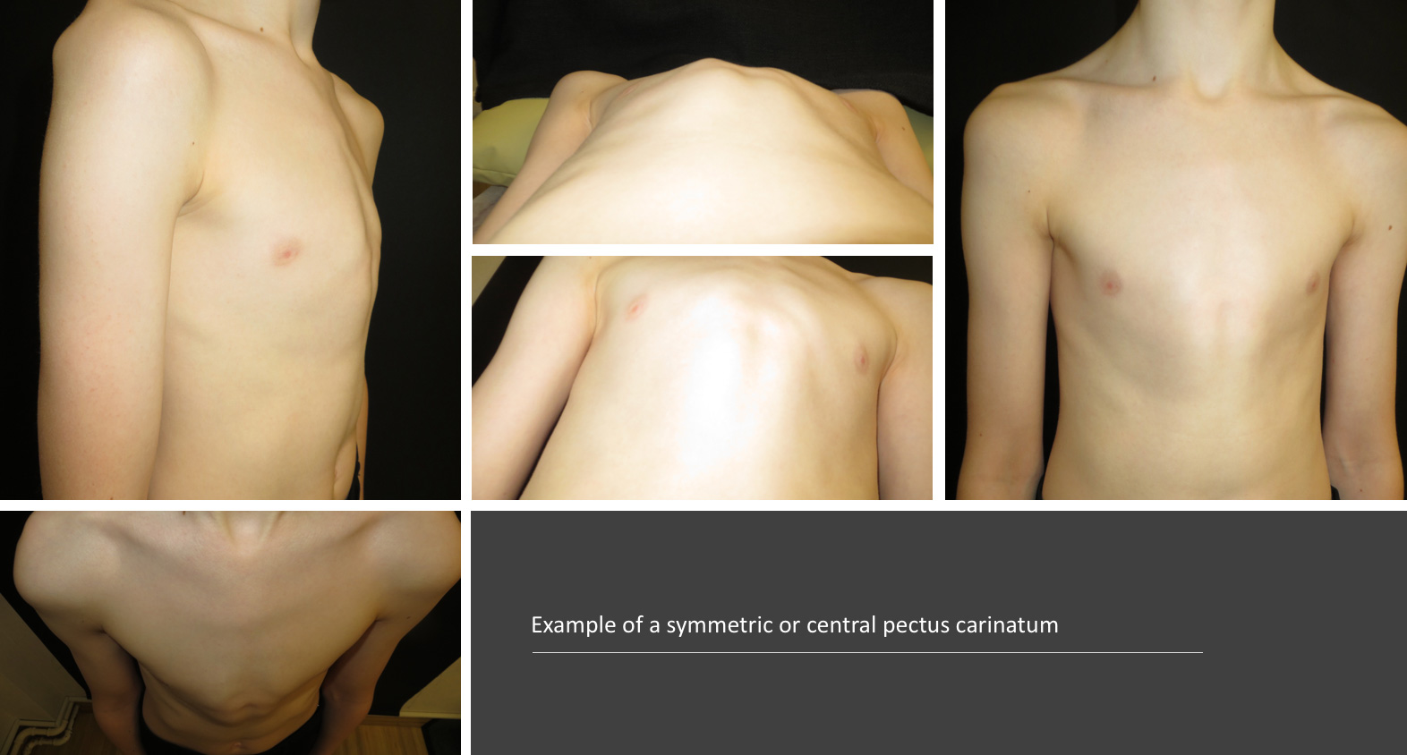
Severe symmetric pectus carinatum deformity in a young boy involving the lower anterior chest wall
Severe symmetric pectus carinatum deformity in a young man with bilateral associated rib flattening
Gallery
We take numerous measurements and photos before, during and after bracing or surgical treatment as many of the problems we see centre around appearance. With the patient's permission we are happy to share our outcomes.
Results
It is important that we can show evidence that bracing or surgical treatment we offer and the results that we achieve are proven. We actively collect data to allow us to study, research and publish are outcomes.
Please click on the buttons below to see and read about patients verified experiences (in their own words) and testimonials (which generally include before and after treatment photos). The pectus clinic is very grateful to all the patients who provided feedback.