Chest wall deformities and rib conditions.
Specialist assessment and treatment for the full range of chest wall deformities — from first presentation through to complex, revision and reconstructive cases — in children, adolescents and adults.
A common condition, often underrecognised.
Chest wall deformities are structural variations of the sternum, costal cartilages and ribs — most arising from altered cartilage growth during development, creating a buckling effect on the chest and sternum. The most familiar are pectus excavatum (sunken chest) and pectus carinatum (pigeon chest), but the range is wide: presentations vary from straightforward single deformities to complex, asymmetric and mixed cases.
Some patients notice changes in their posture before the chest wall shape itself becomes the main concern — often during periods of growth spurts. Rounded shoulders, a forward head position and altered thoracic shape are a recognised part of the clinical picture and are assessed and addressed as part of the overall treatment plan.
Many patients experience significant delay between first noticing their condition and receiving a clear specialist opinion. The initial assessment at the Pectus Clinic is structured to provide a firm diagnosis, a straightforward explanation of the findings, and a treatment plan built around the individual — whether that means reassurance, non-operative management or surgical correction.
Rib flare may occur alongside a pectus deformity or as an isolated finding, and is fully within the scope of the Pectus Clinic. For slipping rib syndrome, rib fractures, costal cartilage injury and chronic chest wall pain, Mr Hunt also leads a dedicated Rib Injury Clinic ↗.


Pectus in numbers
Published literature alongside Pectus Clinic clinical experience and audit data collected through the clinic
Understanding the structures involved
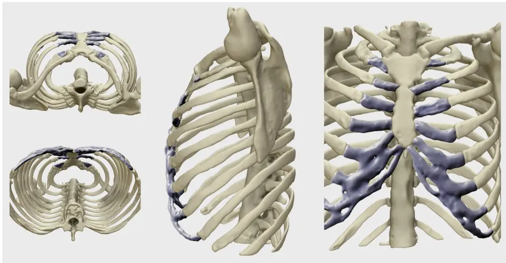
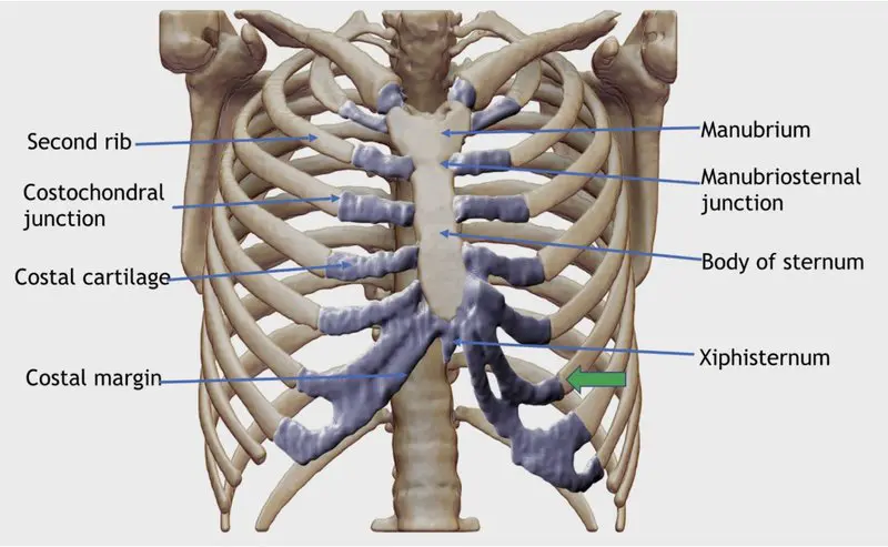
Pectus deformities affect the sternum, costal cartilages and ribs — the structural framework of the anterior chest wall. The costal cartilages connect the ribs to the sternum; altered growth of these cartilages during development is the primary driver of most pectus conditions.
Interactive 3D model — drag to rotate
Normal chest anatomy shown for reference. Rotate and zoom to explore rib, cartilage and sternal relationships.
Annotated anatomy diagrams
Chest wall conditions assessed at the Pectus Clinic.
The clinic assesses and manages a wide range of chest wall deformities and related conditions, from common pectus presentations to complex, asymmetrical and recurrent cases.
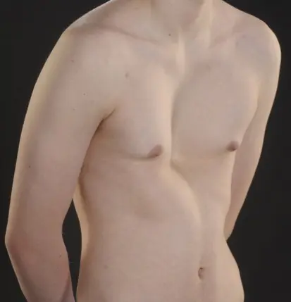
The most common chest wall deformity, in which the sternum and costal cartilages grow inward, creating a concavity in the centre of the chest. Can affect cardiopulmonary function and have a significant impact on body image and quality of life.
Learn more →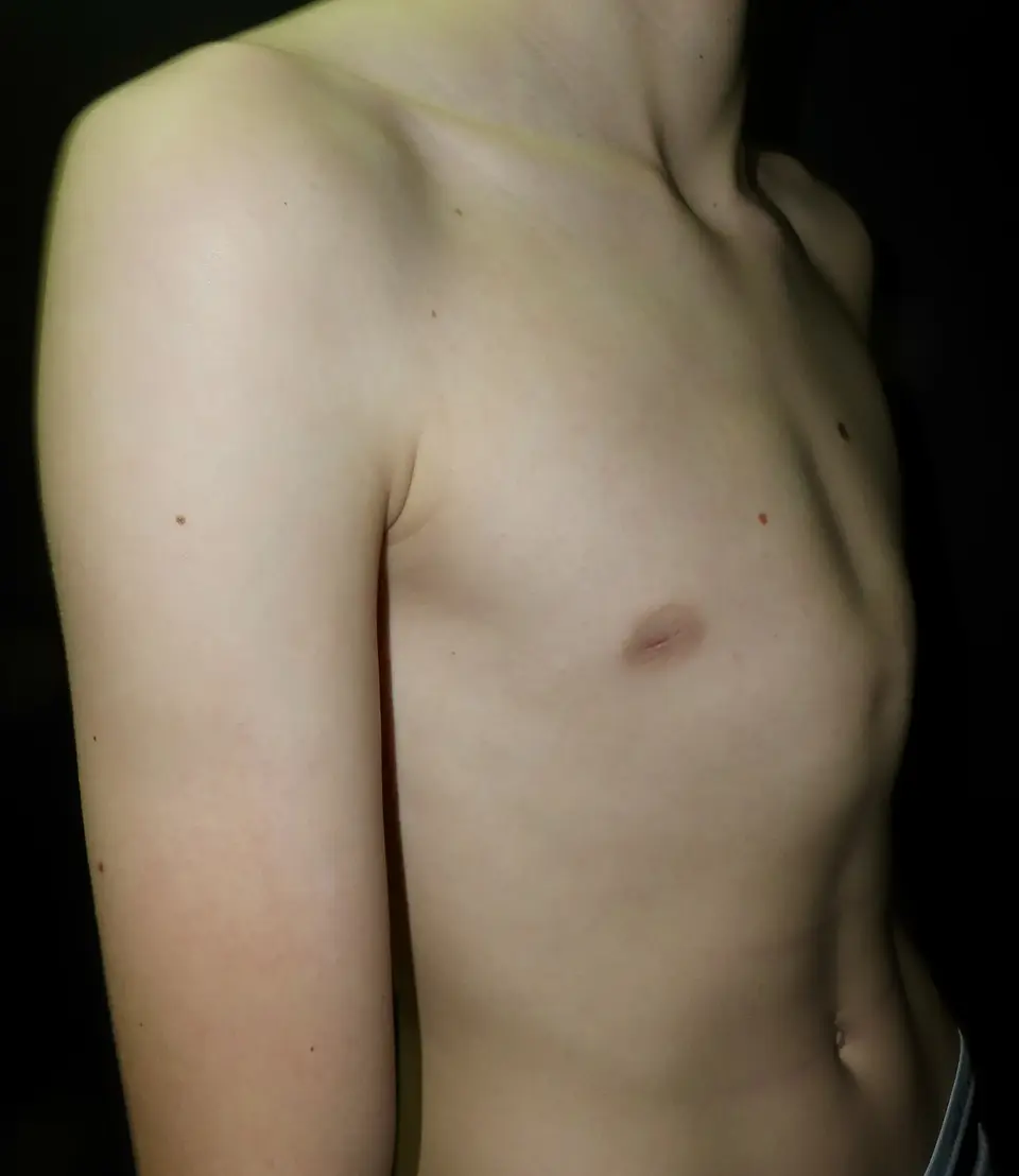
A forward protrusion of the sternum and costal cartilages. Often becomes more noticeable during adolescent growth. Responds well to external compressive bracing in flexible presentations, and may require surgery in rigid or complex cases.
Learn more →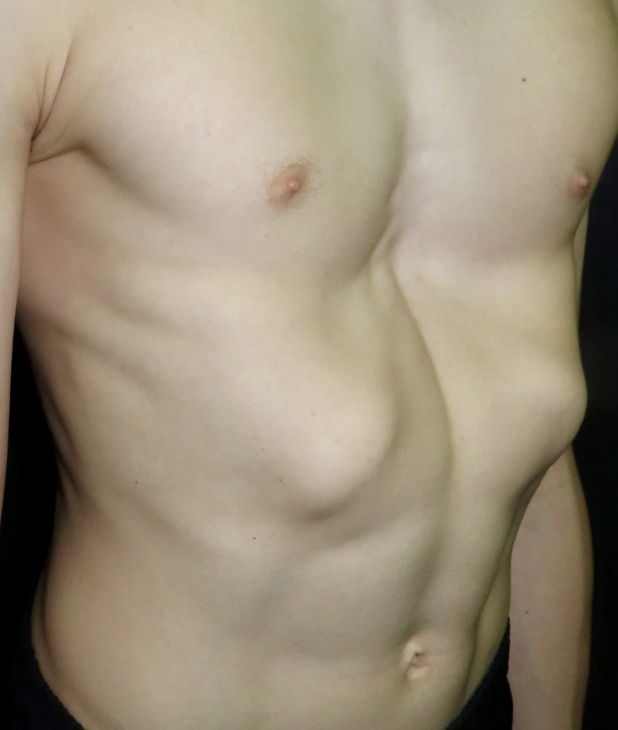
Prominence of the lower ribs — one or both sides — that may occur in isolation or alongside pectus deformities. Present in nearly half of pectus patients. Can cause discomfort, restricted clothing fit and self-consciousness. Specialist bracing and physiotherapy are first-line treatments.
Learn more →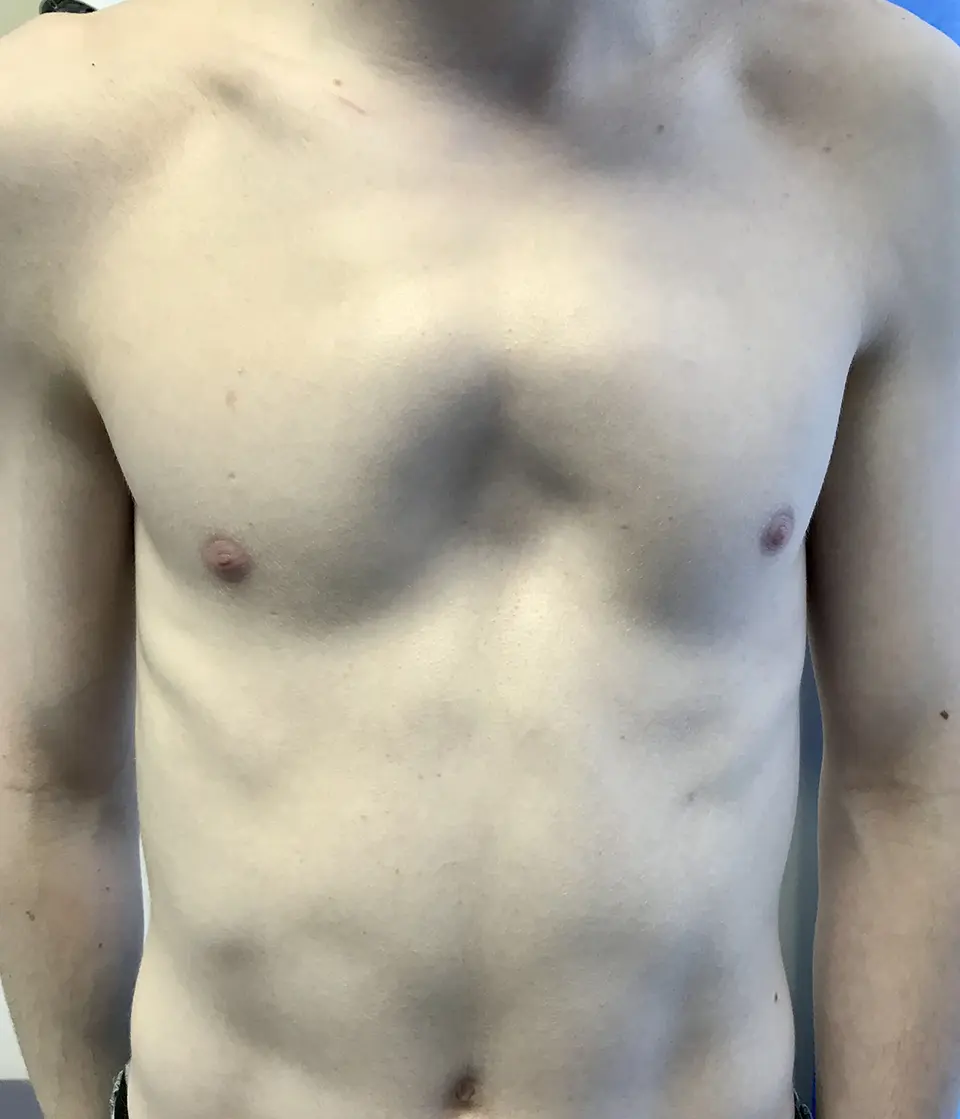
A rare variant in which the upper costal cartilages protrude while the lower sternum may be depressed, creating a complex anterior chest wall shape. Often associated with early fusion of the manubriosternal joint. Requires individualised surgical planning.
Learn more →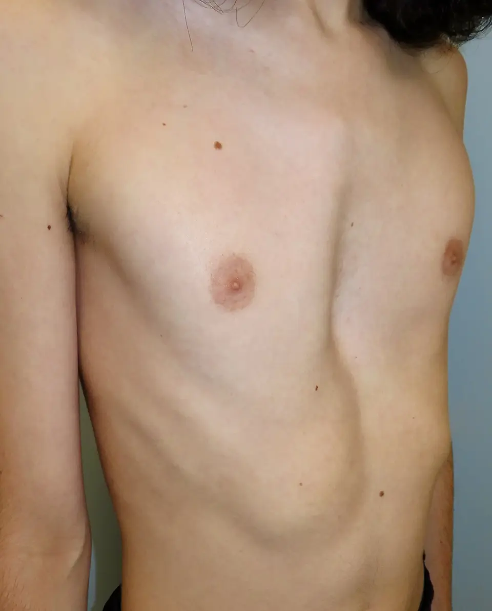
Many patients present with a combination of features — excavatum with carinatum, unilateral deformity, or asymmetric sternal rotation. Horseshoe (Currarino-Silverman) deformity involves protrusion of the upper cartilages with sternal depression. Complex cases require bespoke assessment and operative planning.
Learn more →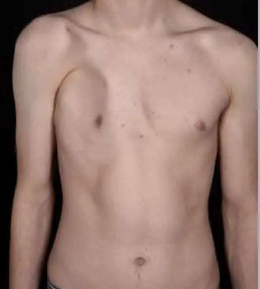
A congenital condition characterised by absence or underdevelopment of the chest wall muscles on one side, often with associated rib and breast anomalies. Chest wall reconstruction — including CAD custom implants and hybrid techniques — can achieve significant improvement in symmetry and contour.
Learn more →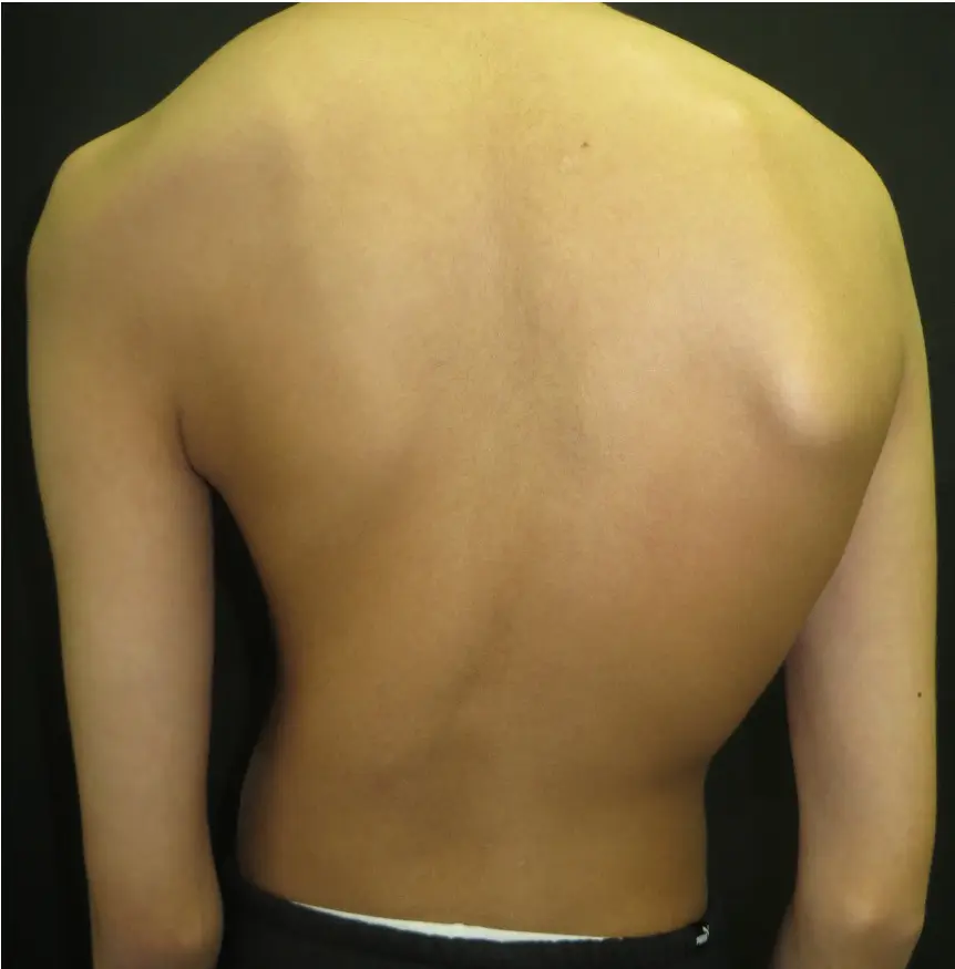
Pectus deformities occur more frequently in association with connective tissue disorders including Marfan syndrome, Ehlers-Danlos syndrome and Noonan syndrome, and with scoliosis. These conditions require careful co-ordination between specialties, with treatment planning tailored to the individual's overall profile.
Learn more →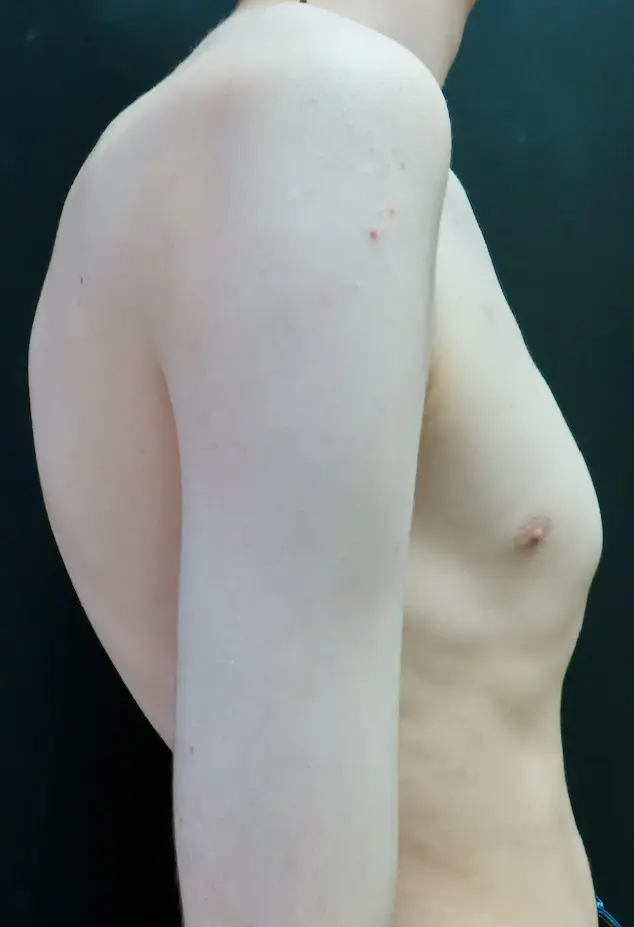
Rounded shoulders, forward head position and altered thoracic shape are frequently seen alongside pectus deformities. Postural changes may be the first thing a patient or parent notices, and are assessed and managed as part of the overall treatment pathway.
Learn more →Not sure which condition applies?
Many patients are uncertain about their diagnosis before they are seen. A structured consultation with Mr Hunt includes a thorough clinical assessment, review of any existing investigations, and a clear explanation of findings — whatever the presentation.