Associated Conditions
Most pectus deformities occur in isolation. In a significant minority, the chest wall abnormality is part of a broader genetic or connective tissue disorder. Identifying these before treatment is planned is essential — both for safety and for optimising outcomes.
When pectus is part of a broader picture
A pectus deformity that forms part of a syndrome or connective tissue disorder requires a fundamentally different approach to assessment and treatment planning compared to an isolated deformity.
In syndromic cases, the chest wall abnormality is one element of a multi-system condition. The connective tissue defect that drives the pectus may also affect the aorta, eyes, joints, skin, and skeletal development. Before any operative intervention is considered, the full picture must be established — including echocardiography, ophthalmological review, and genetic assessment where indicated.
Early recognition also matters because some syndromic patients are managed differently during surgery — altered anaesthetic risk, atypical healing, aortic root dimensions, or bone fragility all affect technique and decision-making. Identifying these factors upfront protects the patient and allows the team to plan comprehensively.
At a glance
Scoliosis
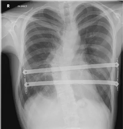
Scoliosis — lateral curvature of the spine — is the condition most frequently associated with pectus excavatum. The two deformities can coexist independently or as part of a shared underlying connective tissue predisposition.
The normal spine has sagittal curves at the cervical and lumbar levels, but runs straight in the coronal plane. In scoliosis, the spine curves sideways in an S or C shape, often with associated vertebral rotation. The severity is graded by Cobb angle on erect AP spine radiograph. Curves below 20° are typically monitored; those between 20–40° may require bracing during growth; curves above 40–50° are usually referred for orthopaedic surgical consideration.
Scoliosis associated with pectus may be congenital (structural, fixed) or non-structural (secondary to muscle imbalance, leg length discrepancy, or post-surgical). The distinction matters clinically: structural scoliosis does not resolve with positional correction, whereas non-structural curves may improve with the underlying cause treated.
When both conditions coexist, imaging must characterise both deformities before any surgery is planned. Combined correction — in selected cases — may be undertaken in a staged or simultaneous approach, coordinated with spinal surgery colleagues.
Clinical features
- Lateral spinal curvature visible on erect AP radiograph; Cobb angle >10° defines scoliosis
- Rib humping on forward flexion (Adam's forward bend test) due to vertebral rotation
- Uneven shoulder height, prominent scapula on convex side, hip asymmetry
- Adolescent idiopathic scoliosis most common — onset typically 10–16 years
- Respiratory compromise in severe curves (>70°) due to thoracic cage distortion
- Reported incidence of scoliosis in pectus excavatum: 15–26% depending on series

Scoliosis Support & Research is the UK charity providing support, information, and advocacy for people affected by scoliosis and related spinal conditions.
Visit SSRS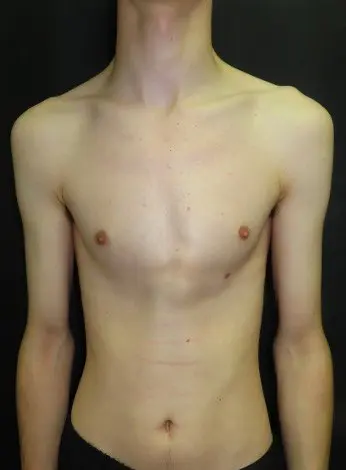
Ghent criteria — key features
- Aortic root dilatation or dissection (major criterion)
- Ectopia lentis — upward lens dislocation (major criterion)
- FBN1 pathogenic variant or positive family history
- Systemic score ≥7 (includes pectus, wrist/thumb signs, scoliosis, flat feet, dural ectasia)
- Pectus carinatum scores 2; pectus excavatum or chest asymmetry scores 1
Marfan syndrome
Marfan syndrome is the commonest connective tissue disorder associated with pectus deformity. It is caused by pathogenic variants in FBN1, the gene encoding fibrillin-1, producing systemic weakness of connective tissue that affects the cardiovascular system, skeleton, and eyes.
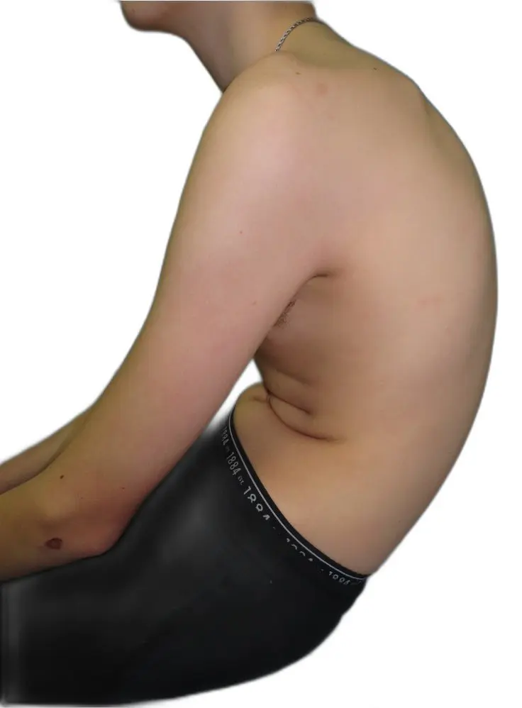
The characteristic skeletal features — tall stature, disproportionately long limbs (dolichostenomelia), long slender fingers (arachnodactyly), and a reduced upper-to-lower segment ratio — are often striking. Pectus deformity occurs in a significant proportion of patients, taking the form of pectus excavatum, pectus carinatum, or asymmetric mixed deformity. The costal cartilages undergo abnormal elongation, driving sternal displacement.
The critical clinical concern in Marfan syndrome is aortic root dilatation, which carries a risk of life-threatening dissection. Before any pectus surgery — including the Nuss procedure — the aortic root dimensions must be known and cardiology clearance obtained. In some cases, aortic root replacement and pectus correction may be planned concomitantly.
Diagnosis is made using the revised Ghent nosology (2010), which requires a combination of aortic criteria, ectopia lentis, systemic features, and genetic testing. All patients presenting with a Marfanoid habitus and pectus should be referred for echocardiography and ophthalmological assessment before treatment is discussed.
Ehlers-Danlos syndrome
The Ehlers-Danlos syndromes (EDS) are a heterogeneous group of inherited connective tissue disorders united by defects in collagen structure or processing. Joint hypermobility, skin hyperextensibility, and tissue fragility are the hallmarks.
Thirteen subtypes are currently recognised, with hypermobile EDS (hEDS) being the most prevalent. In hEDS, pectus excavatum or carinatum may occur as part of the broader musculoskeletal phenotype, alongside joint instability, chronic pain, and autonomic dysfunction. The diagnosis of hEDS currently rests on clinical criteria alone — no causative gene has been identified.
The classical EDS subtypes (caused by collagen V variants) carry a risk of tissue fragility, poor wound healing, and atrophic scarring. These features have direct implications for surgical planning — skin closure technique, wound tension, and recovery expectations all require adjustment in EDS patients. The vascular subtype (vEDS, caused by COL3A1 variants) carries a risk of arterial dissection and represents an absolute contraindication to elective pectus surgery without detailed vascular surgical input.
In patients with known or suspected EDS and pectus, a thorough pre-operative assessment includes evaluation of skin and joint phenotype, wound healing history, and consideration of connective tissue specialist review before any operative decision is made.
Relevant EDS subtypes in pectus practice
- Hypermobile EDS (hEDS): most common; joint laxity, chronic pain, pectus may coexist; surgical risk generally manageable
- Classical EDS (cEDS): fragile skin, poor healing, wide atrophic scarring — requires modified surgical technique
- Kyphoscoliotic EDS (kEDS): progressive scoliosis and muscle hypotonia from birth; significant overlap with pectus deformity
- Vascular EDS (vEDS): arterial and organ rupture risk — elective pectus surgery requires careful multidisciplinary discussion
Screening questions

EDS UK is the national charity supporting people living with Ehlers-Danlos syndrome and hypermobility spectrum disorders — providing information, peer support, and specialist resources.
Visit EDS UKDistinguishing features vs Marfan

Supporting patients and families living with Marfan syndrome, Loeys-Dietz syndrome, and related connective tissue conditions — with information, peer support, and specialist resources.
Visit Marfan TrustLoeys-Dietz syndrome
Loeys-Dietz syndrome (LDS) is a rare autosomal dominant connective tissue disorder caused by pathogenic variants in TGF-β signalling genes. It shares several features with Marfan syndrome but carries a particularly high risk of aggressive aortic and arterial disease.
LDS was first described in 2005 and is caused by variants in genes encoding components of the TGF-β signalling pathway — most commonly TGFBR1 and TGFBR2, though variants in SMAD3, TGFB2, TGFB3, and SKI are also recognised. Five subtypes are described based on genotype.
The skeletal phenotype of LDS overlaps considerably with Marfan syndrome and may include pectus excavatum or carinatum, scoliosis, joint laxity, and tall stature. Distinguishing features include craniofacial abnormalities (hypertelorism, bifid uvula, cleft palate), arterial tortuosity, and the absence of lens dislocation. In some subtypes, skin findings resembling vascular EDS are present.
The vascular risk in LDS is significant and may be more aggressive than in Marfan syndrome — aortic dissection can occur at smaller root diameters. Any patient with a suspected connective tissue disorder and arterial abnormalities on imaging should have LDS considered in the differential before pectus surgery is planned.
Osteogenesis imperfecta
Osteogenesis imperfecta (OI) — brittle bone disease — is a group of genetic bone fragility disorders caused predominantly by pathogenic variants in collagen type I genes (COL1A1, COL1A2). Pectus deformity, though not universal, occurs in a proportion of patients across the OI spectrum.
OI ranges considerably in severity, from type I (mild, compatible with normal lifespan, minimal fracture history) through to type II (perinatal lethal). Types III and IV represent moderate to severe forms with progressive bony deformity. In patients with moderate to severe OI, the thoracic cage may be significantly distorted — rib fractures during infancy and childhood lead to a characteristic bell-shaped thorax with pectus deformity superimposed on abnormal rib architecture.
Clinical features beyond the bones include blue sclerae, dentinogenesis imperfecta, hearing loss, and short stature depending on type. Joint laxity is common in milder forms.
Treatment of pectus in OI requires significant adaptation. External bracing for carinatum carries an uncertain fracture risk in moderate to severe forms. Surgical correction — where indicated — must be undertaken with bone fragility in mind, and standard bar fixation techniques may require modification. OI patients should be managed within an experienced specialist centre with access to metabolic bone and paediatric expertise.
Pectus considerations in OI
- Pectus deformity may reflect rib fracture deformity as well as costal cartilage changes
- CT with 3D reconstruction is essential to understand the skeletal architecture
- External bracing is possible in mild OI but requires caution and monitoring
- Surgical correction requires modified technique — standard sternal retraction forces may cause rib fracture
- Bisphosphonate therapy status must be established pre-operatively
OI classification at a glance
Musculoskeletal features
- Tall, thin stature — Marfanoid habitus
- Pectus excavatum or carinatum
- Long limbs (dolichostenomelia)
- High-arched feet (pes cavus)
- Knock knees (genu valgum)
- Osteoporosis — fracture risk
- Scoliosis in some cases
Distinguishing from Marfan syndrome
Homocystinuria
Homocystinuria is an autosomal recessive inborn error of methionine metabolism caused by deficiency of cystathionine beta-synthase. The resulting accumulation of homocysteine affects connective tissues, the vascular system, the eyes, and cognitive development.
The skeletal phenotype closely resembles Marfan syndrome — tall stature, long limbs, arachnodactyly, and pectus deformity are all well-recognised. The distinction can be made clinically: lens dislocation in homocystinuria is typically downward (rather than the upward dislocation seen in Marfan syndrome), and the vascular risk is thrombotic rather than related to aortic root dilatation. Cognitive impairment of variable degree is common.
In patients on pyridoxine-responsive homocystinuria, skeletal and connective tissue manifestations may be partially attenuated. However, the thromboembolic risk — particularly perioperative — remains significant. Any patient with homocystinuria requiring pectus surgery must have haematological review, pre-operative homocysteine optimisation, and an anti-thrombotic strategy agreed before surgery proceeds.
Noonan syndrome
Noonan syndrome is a relatively common autosomal dominant RASopathy — caused by gain-of-function variants in the RAS/MAPK signalling pathway — and is one of the most frequent genetic causes of congenital heart disease. Pectus deformities are a recognised skeletal feature.
The condition affects approximately 1 in 1,000–2,500 live births. Pathogenic variants in PTPN11 account for roughly half of cases; mutations in SOS1, RAF1, RIT1, and other RAS pathway genes account for most of the remainder. The clinical phenotype is variable even within families.
Pectus deformity in Noonan syndrome may take the form of pectus excavatum, pectus carinatum, or a mixed pattern. The mechanism is likely related to abnormal costal cartilage growth driven by the underlying signalling dysregulation. Scoliosis is also recognised.
The most clinically significant associations are cardiac. Pulmonary valve stenosis is the most common cardiac defect (present in ~50%), followed by hypertrophic cardiomyopathy (~20%) and atrial septal defect. Bleeding disorders — due to platelet dysfunction and clotting factor deficiencies — are present in a significant minority and must be evaluated pre-operatively. Short stature and mild-to-moderate learning difficulties complete the phenotype in many patients.
Key clinical features
- Facial features: widely spaced eyes (hypertelorism), ptosis, low-set posteriorly rotated ears, short neck with webbing, small chin
- Cardiac: pulmonary valve stenosis, hypertrophic cardiomyopathy, ASD
- Skeletal: pectus excavatum or carinatum, scoliosis, short stature
- Haematological: platelet dysfunction, factor XI deficiency — bleeding risk
- Skin: café-au-lait spots, lymphoedema in some cases
- Increased risk of lymphoma and leukaemia
Pre-operative considerations
Turner syndrome
Turner syndrome — caused by complete or partial monosomy of the X chromosome (45,X or mosaic variants) — affects approximately 1 in 2,500 female births and carries well-recognised cardiac, skeletal, and endocrine associations. Chest wall abnormality is a recognised feature that may present to a pectus clinic.
The characteristic chest wall finding is a broad, shield-shaped thorax with wide-spaced nipples. Pectus excavatum or pectus carinatum may coexist and can be the presenting complaint in patients whose Turner syndrome has not yet been diagnosed. Short stature, webbed neck (pterygium colli), low posterior hairline, and primary amenorrhoea complete the classic phenotype, though the presentation is highly variable — particularly in mosaic forms.
The critical clinical concern is cardiovascular. Bicuspid aortic valve occurs in 15–30% of Turner patients, and coarctation of the aorta in approximately 10%. Aortic root dilatation and dissection — though less common than in Marfan syndrome — are recognised risks, occurring at smaller aortic dimensions relative to body surface area. Cardiovascular assessment, including echocardiography and cardiac MRI, is mandatory before any pectus surgery is considered.
Osteoporosis is common due to oestrogen deficiency and represents an additional surgical consideration — bone quality must be assessed if implant fixation or sternal correction is planned. Growth hormone treatment and oestrogen replacement therapy must be documented and reviewed in the pre-operative work-up.
Key features and surgical considerations
- Chest wall: broad shield chest, pectus excavatum or carinatum, wide-spaced nipples
- Cardiac: bicuspid aortic valve (15–30%), coarctation of the aorta (~10%), aortic root dilatation — echo and cardiac MRI mandatory pre-operatively
- Skeletal: short stature, cubitus valgus, osteoporosis — bone quality relevant for fixation
- Endocrine: oestrogen deficiency, growth hormone treatment — document current therapy
- Lymphoedema: congenital or acquired — may affect wound healing and post-operative recovery
- Renal anomalies (horseshoe kidney) in ~30% — relevant for pre-operative imaging review
Pre-operative checklist
Common causes
Assessment requirements
- Full operative history — previous sternotomy, implants, patches, or vascular grafts
- CT with 3D reconstruction to characterise current anatomy and surgical access
- Cardiology review in post-cardiac surgery patients — residual haemodynamic status
- Anaesthetic review — post-sternotomy patients may have pericardial adhesions
- Cardiothoracic surgical liaison where re-sternotomy risk exists
Post-surgical chest wall deformity
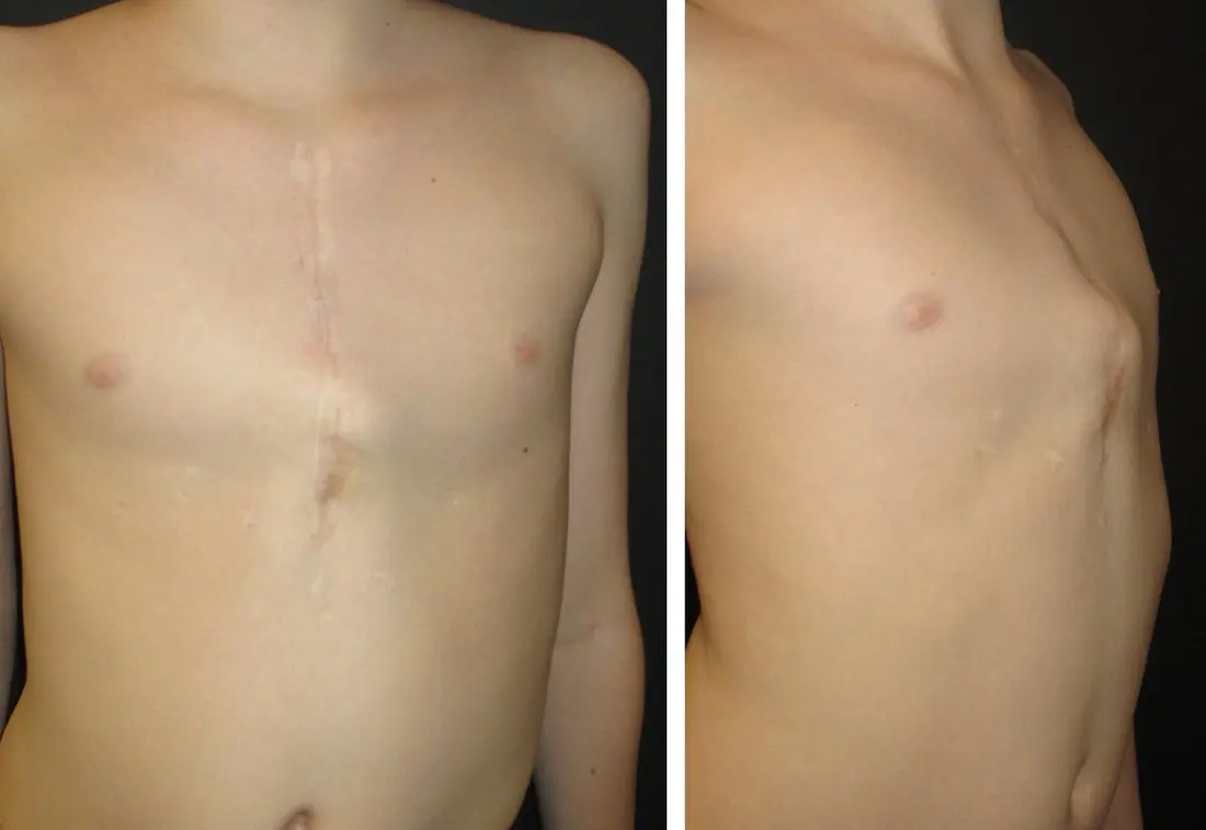
Chest wall deformity arising after earlier surgery represents a clinically distinct category from primary pectus deformity. The surgical anatomy is altered, the cause is known, and the approach to correction requires careful individual planning.
The most common cause seen in pectus practice is chest wall deformity following childhood cardiac surgery via median sternotomy. Disruption of the sternum and anterior chest wall during a period of active skeletal growth — particularly in infancy and early childhood — can result in sternal malunion, asymmetric growth of the costal cartilages, and progressive pectus-type deformity as the patient grows. The resulting deformity may resemble pectus excavatum, pectus carinatum, or a complex asymmetric pattern.
Congenital diaphragmatic hernia (CDH) repair in the neonatal period is another recognised cause. The combination of underlying lung hypoplasia, thoracic cage compression, and surgical repair produces progressive chest wall asymmetry and scoliosis in a proportion of survivors. As CDH survival rates improve, the number of patients reaching adolescence and adulthood with significant chest wall deformity is increasing.
Oesophageal atresia and tracheo-oesophageal fistula (TOF) repair — typically performed via right thoracotomy — may produce chest wall asymmetry, scoliosis, and rib crowding on the approach side. Radiotherapy to the chest in childhood, given for tumours such as Wilms' tumour or neuroblastoma, can cause asymmetric growth plate injury with resultant chest wall distortion.
In all post-surgical cases, the existing surgical history must be fully documented before any new operative procedure is planned. Previous implants, prosthetic patches, vascular grafts, and pericardial adhesions all affect surgical access and risk. CT with 3D reconstruction is essential to characterise the current anatomy.
Cardiac associations
Pectus deformities — both isolated and syndromic — carry recognised associations with cardiac abnormalities. Understanding these associations is important for pre-operative screening and anaesthetic planning.
The relationship between pectus and cardiac abnormalities is not always causal. In some cases — particularly mitral valve prolapse in excavatum — the anatomical distortion of the thorax may directly displace or distort cardiac structures. In others, a shared connective tissue predisposition underlies both the chest wall and cardiac abnormality.
Mitral valve prolapse
Mitral valve prolapse is reported more frequently in patients with pectus excavatum than in the general population, although published rates vary widely depending on age group, diagnostic criteria and study design. Older series reported rates of around 15–65%, while more recent studies commonly report rates in the region of 25–40%. The association is likely multifactorial, reflecting both altered cardiac position and compression from the depressed sternum and, in some patients, an underlying connective tissue predisposition. Most cases are mild and haemodynamically insignificant, but echocardiography is commonly used in pre-operative assessment to evaluate valve function, cardiac compression and associated abnormalities.
Aortic root dilatation
Aortic root dilatation is an important finding in patients with syndromic pectus deformity, particularly in conditions such as Marfan syndrome and Loeys-Dietz syndrome. It should be actively assessed in patients with a Marfanoid habitus, positive family history, or other features of connective tissue disorder. In these patients, echocardiography is used to measure the aortic root and assess associated valve abnormalities, with CT or MRI considered when broader aortic imaging is required. Unrecognised aortic root dilatation may indicate an underlying heritable thoracic aortic disease and can carry a significant risk of progressive aneurysm or dissection, particularly if surgical stress, hypertension or delayed diagnosis are not addressed.
Right heart compression
In severe pectus excavatum, the depressed sternum may directly compress the right ventricle, reducing cardiac output during exertion. This is a well-recognised mechanism of exercise intolerance in moderate-to-severe PE and is objectively demonstrable on cardiac MRI or echocardiogram. Correction of the deformity reliably resolves this finding in the majority of patients.
Congenital heart disease
Congenital heart disease is reported in a small but increased proportion of patients with pectus deformity compared with the general population. Published series vary, but CHD has been reported in approximately 1.5–2% of pectus patients, compared with a background prevalence of around 0.8% in the general population. Most detected abnormalities are minor, but clinically significant lesions can occasionally be identified. For this reason, echocardiography is commonly included in pre-operative assessment, particularly in patients with severe deformity, symptoms, abnormal clinical findings, syndromic features, or a family history of cardiac disease.
Poland syndrome
A distinct congenital chest wall condition
Poland syndrome — characterised by unilateral absence or hypoplasia of the pectoralis major, with variable rib, costal cartilage, and hand involvement — is classified separately from the connective tissue syndromes. It is not caused by costal cartilage overgrowth but by a primary developmental failure of the chest wall musculoskeletal structures, arising during the sixth week of embryogenesis.
It may co-exist with a pectus deformity and may be associated with breast asymmetry in female patients. The full spectrum — from isolated pectoral hypoplasia through to absent ribs with chest wall herniation — requires characterisation with CT imaging before any surgical planning.
Recognising associated conditions in clinical practice
A structured clinical assessment identifies the majority of significant associations before any treatment is planned. Key red flags include a Marfanoid habitus, positive family history, joint hypermobility, scoliosis, or aortic root abnormality on imaging.
Concerned about an associated condition?
If you or your child has a pectus deformity alongside features of a connective tissue disorder, scoliosis, or cardiac abnormality, a specialist assessment will clarify the diagnosis and guide safe, appropriately sequenced treatment.