
Pectus Excavatum
Sunken Chest
The most common chest wall deformity, caused by abnormal growth of the costal cartilages, pushing the sternum inwards. It ranges from mild and barely noticeable to severe, with significant physical and psychological impact.

What is pectus excavatum?
Pectus excavatum — from the Latin meaning "hollowed chest" — is a structural deformity in which the breastbone (sternum) and several rib cartilages grow inwards, creating a concave or sunken appearance to the front of the chest wall.
The deformity may be present at birth, but typically develops later becoming more apparent during adolescent growth spurts. It results from abnormal overgrowth of the costal cartilages — the flexible junctions between the ribs and the sternum — which push the lower sternum backwards rather than allowing the chest to expand outwards normally.
In mild forms, pectus excavatum may cause little more than a cosmetic concern. In more severe cases, particularly where the sternum is displaced significantly or rotated asymmetrically, it can compress the heart and lungs, leading to measurable functional impairment alongside the well-documented psychological burden.
Key facts — Pectus Excavatum
3D anatomy — pectus excavatum chest wall
Interactive 3D model — click and drag to rotate. Use scroll to zoom. Powered by Sketchfab.
Physical symptoms
Symptoms vary widely between individuals and do not always correlate directly with the severity of the deformity. Many patients have significant complaints with moderate-looking excavatum; others with deep deformities may report minimal symptoms. When present, symptoms are typically functional — meaning they arise during or after physical activity rather than at rest — and include breathlessness on exertion, reduced exercise tolerance, and a sense of chest tightness or inability to take a full breath during sport or sustained effort.
Breathlessness on exertion
A common complaint, particularly during exercise, sport or sustained physical effort. Standard lung function (spirometry) is usually normal, but more sensitive cardiopulmonary exercise testing (CPET) may demonstrate reduced oxygen uptake and ventilatory reserve — evidence of real physiological limitation that responds to surgical correction in selected cases.
Exercise intolerance
Reduced stamina, earlier fatigue and poor athletic performance compared to peers — a common trigger for initial presentation. In severe cases, measurable reductions in cardiac output on exercise have been documented, thought to result from the sternum impinging on the right ventricle, reducing stroke volume.
Palpitations
Awareness of irregular or rapid heartbeat, more common in severe deformities where the heart is mechanically compressed and displaced to the left. Some patients have documented arrhythmias, particularly mitral valve prolapse — found in up to 65% of patients with Marfan-associated pectus.
Pre-syncope & dizziness
In severe forms, particularly during vigorous exercise, some patients experience episodes of light-headedness or fainting (syncope). This is rare but clinically important — it is thought to result from dynamic compression of the heart by the indrawn sternum during high-demand states, reducing cardiac output transiently. Always warrants thorough cardiological assessment.
Chest & back pain
Aching or sharp pain across the sternum and anterior chest wall, often with referred discomfort into the mid-back. Typically musculoskeletal in origin. Worse during growth spurts, after exercise, or with sustained postures. Often dismissed as non-specific and may have been present for years before diagnosis.
Poor posture
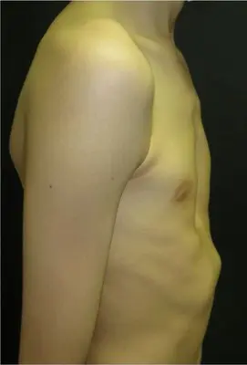
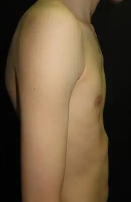
Rounded shoulders, a sunken chest and a forward-curved upper spine (kyphosis) are extremely common and often precede formal diagnosis. Poor posture is not always simply behavioural — it frequently reflects the chest wall shape, with the body compensating for the concavity. Physiotherapy can help significantly but the postural pattern often returns without treatment of the underlying deformity.
Psychological impact
For many patients — particularly adolescents — the psychological burden of pectus excavatum may be as significant as any physical symptom. The visible deformity affects body image at a developmentally vulnerable time, with consequences that can last well into adulthood if not addressed.
Avoidance behaviours are very common: patients routinely avoid swimming pools, changing rooms, beaches, sports, relationships and any situation where the chest might be exposed. Social withdrawal, anxiety and low self-esteem are well documented in the literature and are recognised as valid indications for treatment, independent of objective physiological severity.
The Pectus Clinic takes psychological impact seriously as part of the clinical assessment. Referral to specialist psychological support is available where needed, both as a standalone intervention and as part of the surgical pathway.
How severity and shape vary
Pectus excavatum exists on a wide spectrum and may be described clinically as mild, moderate, severe or extreme. Severity is most commonly graded radiologically using the Haller Index on CT scanning, but the clinical equivalent is the external sternal depression depth, measured as the visible depth of the chest wall hollow. Pectus excavatum can also be described by its morphology. Common patterns include a deep localised cup-shaped depression, a broader shallow saucer-shaped deformity, an elongated trench-like depression, and asymmetric forms associated with sternal rotation, chest wall imbalance or pectoral muscle asymmetry. Assessment of the shape, symmetry, sternal depth, rib flare and posture is important because two patients with similar Haller Indices may have quite different chest wall shapes and treatment considerations. No two cases are identical.
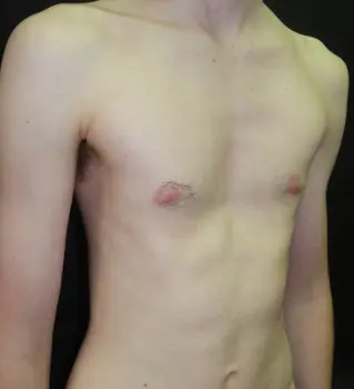
Mild symmetrical — saucer-shaped
A broad, shallow central depression in which the sternum is gently indrawn across a wide area. Symmetrical with no significant rib flaring. Often the least visually conspicuous form — symptoms, if present, are predominantly psychological. Haller Index typically 2.5–3.0.
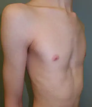
Mild symmetrical — cup-shaped
A focal, circular depression centred over the lower sternum, with the chest wall otherwise symmetrical. Common in adolescence. May be asymptomatic or associated with mild exercise-related awareness. Haller Index typically 2.5–3.2.
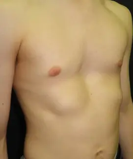
Moderate symmetrical — saucer-shaped
A broader and deeper saucer depression extending across the lower two-thirds of the sternum, with more costal cartilage involvement. Often associated with exercise-related breathlessness and psychological impact. Haller Index typically 3.0–3.5.
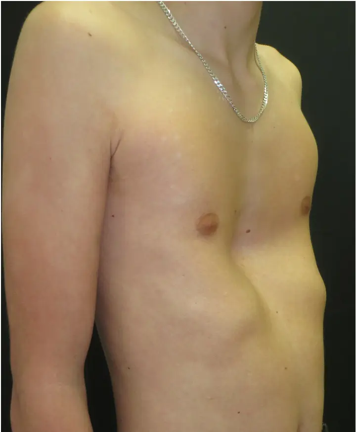
Moderate asymmetric — with rib flaring
Cup or saucer depression combined with visible lower costal margin flaring. The rib flare reflects compensatory chest wall remodelling and is often an independent cosmetic concern. Both the central depression and the peripheral flaring must be assessed in any treatment plan.
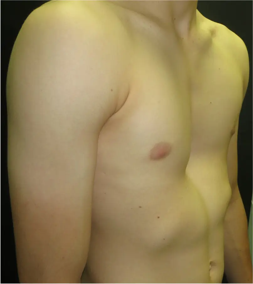
Severe asymmetric — cup-shaped
Deep focal depression with sternal tilt or rotation, resulting from unequal costal cartilage overgrowth. Cardiac displacement towards the left is typical and may be demonstrable on CT and on formal cardiopulmonary exercise testing. Haller Index typically >3.5. Surgical planning must account for the rotational component.
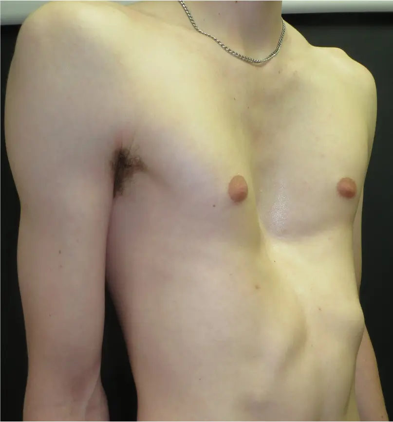
Severe symmetric — trench-shaped
An elongated, canal-like depression running vertically down the sternum, reflecting diffuse costal cartilage involvement. Symmetrical with limited sternal rotation. The length and depth of the trench requires careful preoperative planning. Haller Index typically >3.5.
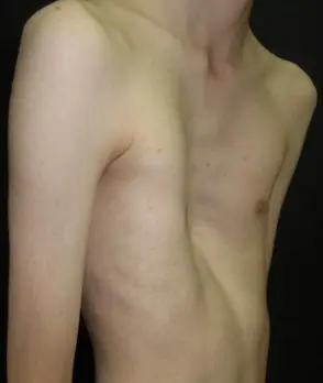
Extreme symmetric — Canal-type / Grand Canyon type
A very deep elongated trench with marked posterior sternal displacement and significant loss of anterior chest wall volume. Cardiorespiratory impairment is likely and demonstrable on formal testing. Haller Index typically >4.5. Detailed multidisciplinary preoperative planning is essential.
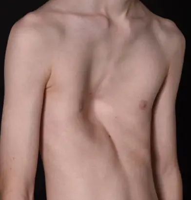
Extreme asymmetric — trench-like
Profoundly deep, asymmetric deformity with significant sternal rotation and chest wall imbalance. Typically the most functionally compromising presentation, with marked compression of the right ventricle and pericardium. A tailored surgical strategy — a multi-bar Nuss, hybrid or even a modified Ravitch in much older patients — is required.
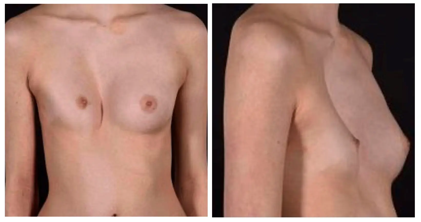
Female presentation
In female patients, breast development may partially conceal the underlying deformity, complicate external measurement and add significantly to the psychological burden. Breast asymmetry — a common associated finding — must be carefully factored into surgical planning, particularly when considering the Nuss procedure or a pectus implant. Three-dimensional imaging and close liaison with a plastic or breast surgeon may be required in selected cases.
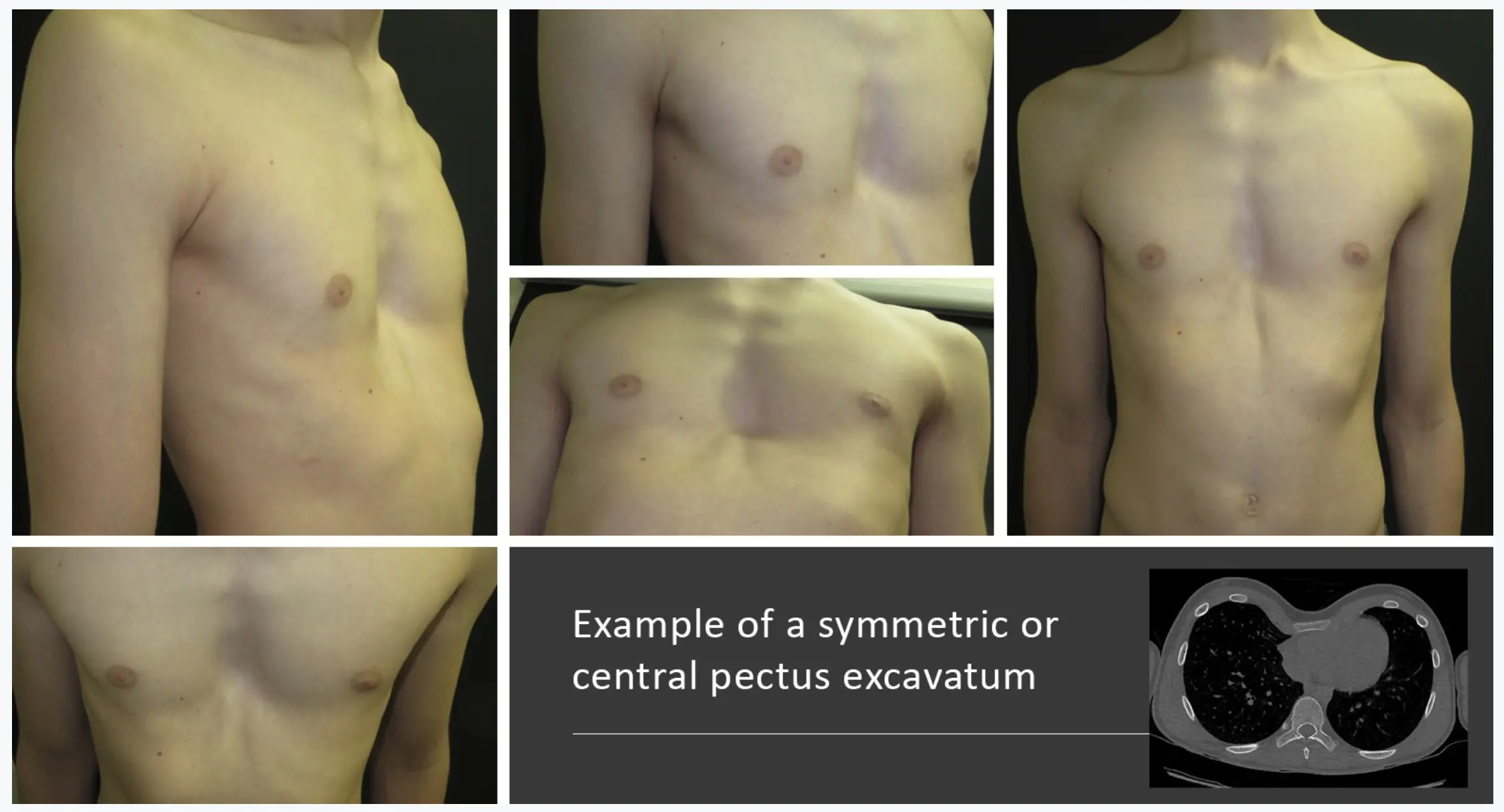
Symmetric (central) excavatum
The depression is centred over the midline with roughly equal depth on both sides. This is the most common pattern and the most straightforward to assess and treat. The sternum sits centrally and does not rotate significantly.
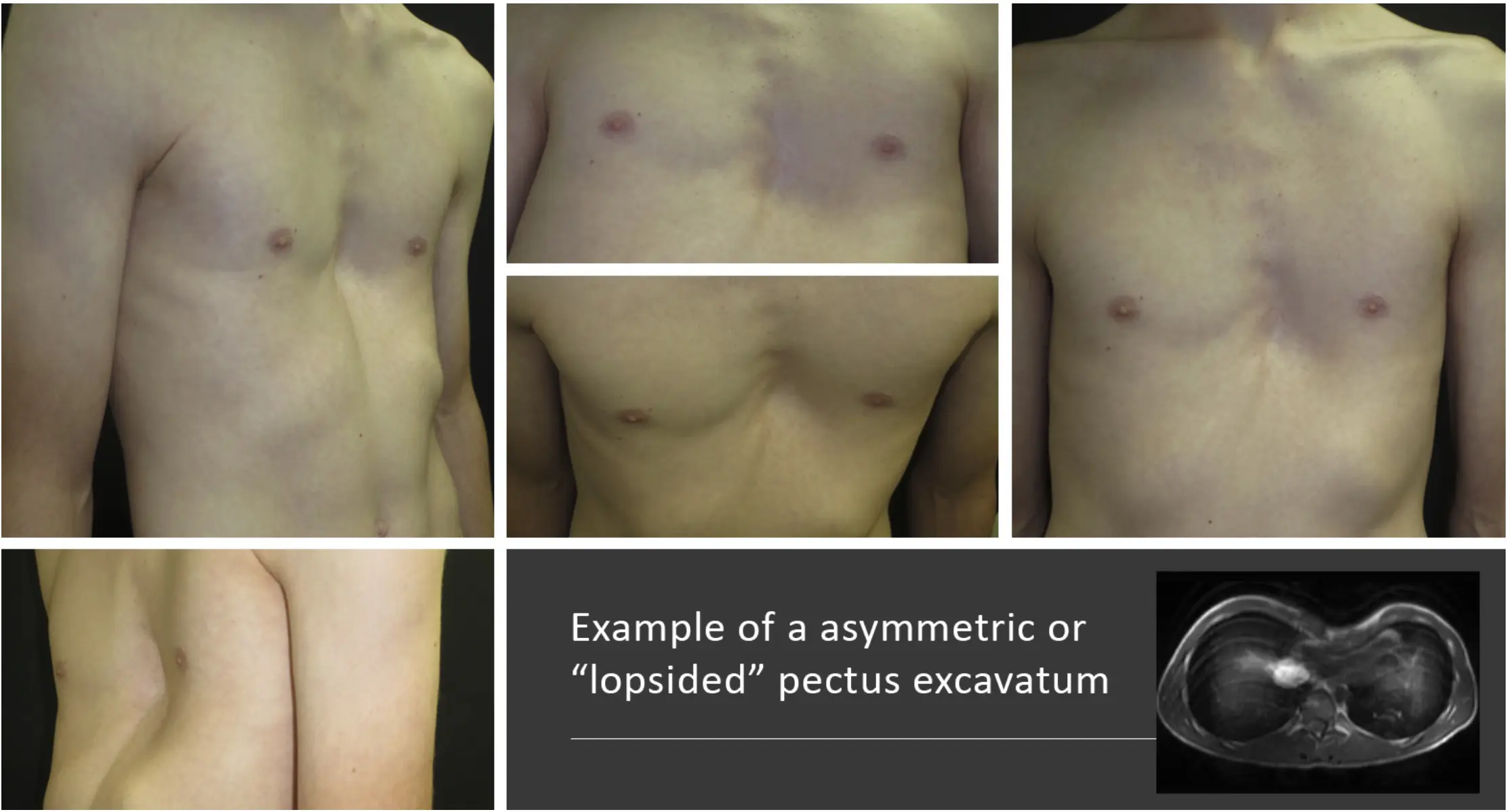
Asymmetric (lopsided) excavatum
The depression is more prominent on one side — typically the right — with the sternum rotating and tilting. This creates an uneven chest profile that can be more complex to correct surgically. The degree of rotation is a key factor in choosing between Nuss and open (Ravitch/hybrid) repair.
How pectus excavatum is assessed
Pectus excavatum is a clinical diagnosis — careful examination by an experienced specialist is usually sufficient. However, structured investigation is important for planning treatment, confirming severity, and identifying associated conditions.
Clinical assessment
Detailed inspection and measurement of the deformity including depth, width, symmetry, sternal rotation, rib flare and posture. Photographs and surface measurements are recorded at baseline. No specific blood test is required for diagnosis.
CT chest (Haller Index)
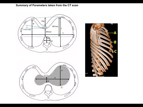
The most useful radiological investigation. A cross-sectional CT image through the deepest point of the deformity allows calculation of the Haller Index — the standard measure of pectus severity. Also identifies sternal rotation, heart displacement, scoliosis and any pulmonary compression. Recommended before surgery.
MRI chest
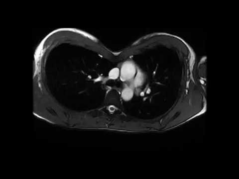
A radiation-free alternative to CT for calculating Haller Index and assessing heart and lung position, but does not provide the same level of radiological detail. In more complex deformity, a CT may be recommended. Preferred in children and younger adolescents with straightforward pectus excavatum, or where repeated imaging may be needed, as it avoids ionising radiation exposure.
Cardiopulmonary exercise test (CPET)
Standard spirometry is usually normal in pectus excavatum. CPET — which measures oxygen consumption and ventilatory function during graded exercise — is a far more sensitive tool and can demonstrate meaningful physiological impairment. Increasingly important in the surgical consent discussion and in post-operative outcome data.
Echocardiogram
Not routinely required, but indicated where symptoms suggest cardiac compression, arrhythmia, or when an associated connective tissue disorder (Marfan syndrome, EDS) is suspected. Mitral valve prolapse is found in up to 65% of Marfan-related pectus cases. Dynamic echocardiography during exercise can demonstrate functional cardiac compromise in severe deformities. Note: pectus deformity can make echocardiogram studies technically challenging and these are best performed by an experienced specialist.
Cardiac MRI
Cardiac MRI provides the most detailed non-invasive assessment of cardiac structure, function and haemodynamics in the context of chest wall deformity. It can quantify ventricular volumes, ejection fraction, stroke volume and the dynamic effects of the deformity on cardiac filling during respiration — information that echocardiography may not reliably capture when image quality is limited by the chest wall shape. When requested in the context of pectus excavatum, it is important that the study is performed as a dedicated pectus protocol by cardiac imaging specialists who are familiar with the condition and its effects on cardiac anatomy.
Genetic assessment
Where Marfan syndrome, Ehlers-Danlos syndrome or another connective tissue disorder is suspected clinically, genetic assessment and ophthalmological review should be arranged before any surgical intervention. Aortic root dimensions must be assessed in Marfan-associated cases before the Nuss procedure is considered.
When surgery is being considered, a CT scan is taken at the deepest point of the depression. Two measurements are calculated from this image to quantify the severity of the deformity.
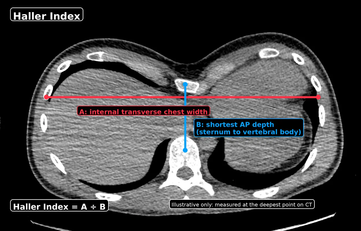
Haller Index = chest width (a) ÷ chest depth (b)
The Haller Index compares how wide the chest is to how much front-to-back space exists behind the sternum. Because the sternum is pushed inwards in pectus excavatum, the front-to-back space is reduced — making the Haller Index higher. The deeper and narrower the deformity, the higher the number.
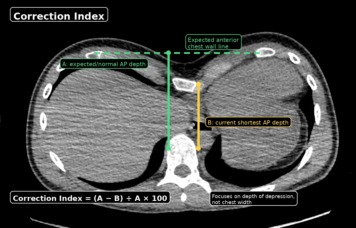
Correction Index = (expected depth − actual depth) ÷ expected depth × 100
The Correction Index expresses the depth of the sternal depression as a percentage of the expected chest depth — in other words, how much of the available chest space has been lost. It is useful because it does not depend on chest width, making it helpful in patients with narrow or unusually shaped chests. A value above 30% is generally considered significant.
The Haller and Correction Indices are useful guides, but they do not capture everything. Two patients with the same index may have very different chest shapes, symptoms and treatment needs. Measurements are always interpreted alongside clinical examination, symptoms and patient goals.
Conditions commonly seen alongside pectus excavatum
Pectus excavatum rarely exists in isolation. Careful assessment should always include screening for the following, which can influence treatment selection and timing.
Rib flare
The lower costal margin (bottom of the rib cage) protrudes outwards as a direct consequence of the indrawn sternum. Can be a significant cosmetic concern in its own right and may worsen during vacuum bell therapy in younger patients. Learn about rib flare →
Poor posture (pectus posture)
Rounded shoulders, kyphosis and forward head carriage are almost universal in pectus excavatum. Physiotherapy can address the postural component independently, but the underlying deformity often limits the extent of correction achievable without treatment. Physiotherapists experienced in pectus-related posture are available through the clinic's specialist network. Learn about pectus posture →
Scoliosis
Lateral spinal curvature is found in approximately 15–20% of patients with pectus excavatum. Significant scoliosis may influence surgical approach — specifically whether a Nuss bar can be placed safely — and should be assessed with plain radiographs or CT in surgical candidates. Where scoliosis requires further evaluation or treatment, referral to a specialist spinal team is coordinated as part of the assessment pathway.
Marfan syndrome
Pectus excavatum is one of the cardinal features of Marfan syndrome, a heritable connective tissue disorder. Key concerns include aortic root dilatation and lens dislocation. Any patient with tall stature, arm span > height, or family history of sudden cardiac death warrants genetic and cardiology review before surgery. Coordinated assessment through a multidisciplinary specialist network — including cardiology, genetics and ophthalmology — is arranged where Marfan syndrome is suspected or confirmed.
Ehlers-Danlos & hypermobility
Generalised joint hypermobility and Ehlers-Danlos syndrome (particularly the hypermobile subtype) are more prevalent in patients with chest wall deformities. This can affect pectus posture, pain patterns and the response to physical therapy. Pain management strategies may need to be adjusted accordingly. Where specialist input is needed, referral to rheumatology or physiotherapists experienced in hypermobility-related chest wall problems can be arranged through the clinic network.
Poland syndrome
A rare condition involving underdevelopment of the pectoralis muscle and, in some cases, breast and rib abnormalities on one side. When pectus excavatum occurs in the context of Poland syndrome it is typically asymmetric and requires tailored reconstructive planning. Liaison with plastic surgery and specialist breast reconstruction colleagues is coordinated through the clinic where required. Learn about Poland syndrome →
Treatment options for pectus excavatum
There is no single right treatment for pectus excavatum. The most appropriate approach depends on age, severity, deformity type, symptom burden, patient priorities and clinical assessment. Mr Hunt offers the full range of treatments and will recommend the most suitable option at consultation.
Vacuum Bell Therapy (VBT)
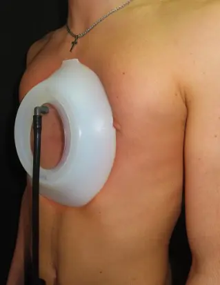
A non-surgical device worn for approximately 4 hours daily over 2 years that lifts the sternum using gentle suction. Developed by Eckart Klobe, it is the only established non-surgical treatment for pectus excavatum. Works best in younger patients with flexible chest walls. Requires commitment and regular specialist follow-up.
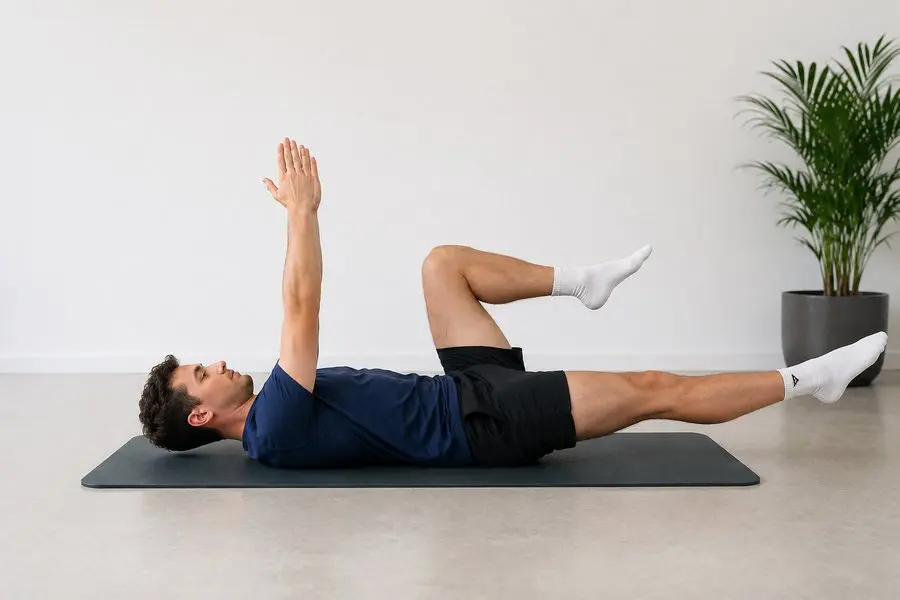
Physiotherapy & Posture Correction
Targeted chest wall exercises, breathing techniques and postural training to strengthen the chest and upper back musculature, reduce kyphosis and improve confidence. Cannot correct the underlying deformity but can significantly improve symptoms, posture and function — both independently and as an adjunct to other treatments.

Psychological Support
For many patients the psychological impact is the primary concern. Access to specialist psychological support — including cognitive behavioural therapy, body image work and acceptance-based approaches — is an important part of the care pathway. Available independently of and alongside physical treatments.
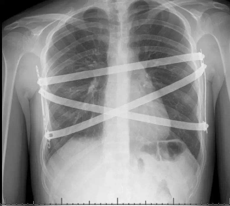
Nuss Procedure
The minimally invasive standard for pectus excavatum repair. Two or more curved titanium bars are inserted through small lateral incisions and flipped behind the sternum to push it outward and secured to the chest wall. No cartilage is removed. The bars remain in place for 3–4 years while the chest remodels, then are removed under a second general anaesthetic. Excellent long-term outcomes in appropriately selected patients.
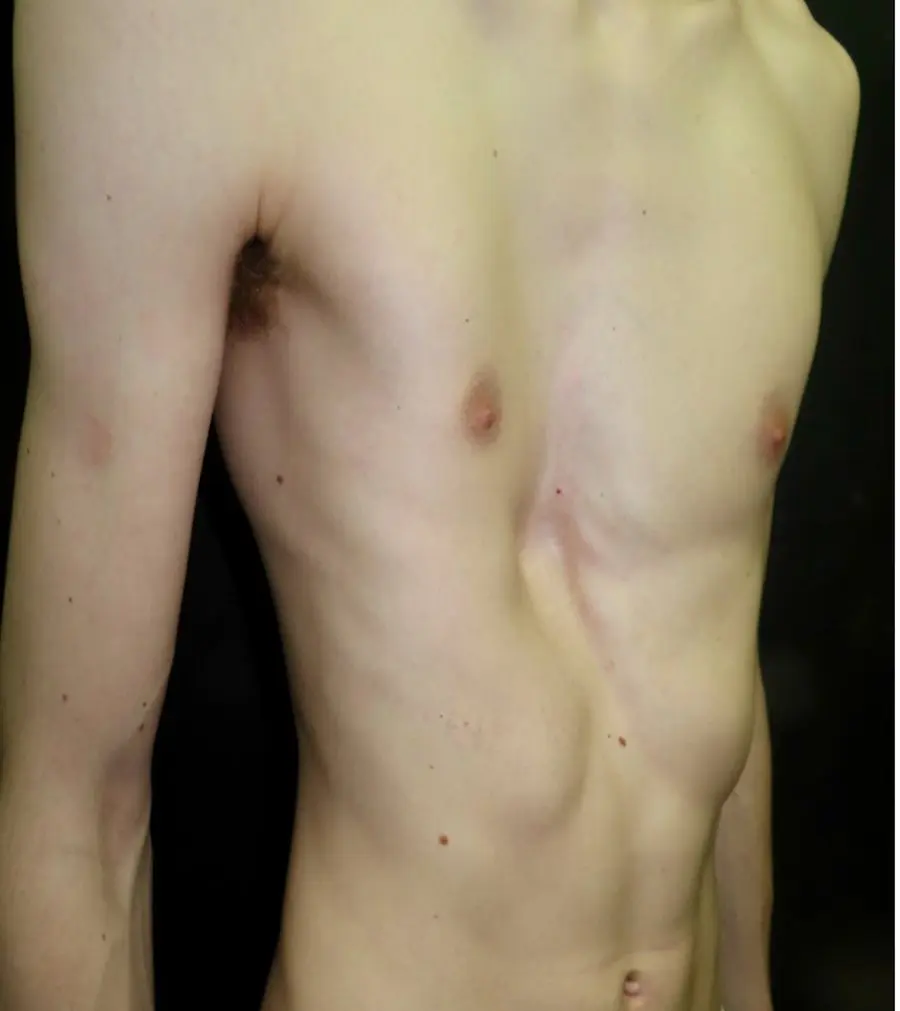
Ravitch / Hybrid Procedure
An open surgical repair in which the abnormal costal cartilages are divided or partially removed, the sternum is repositioned, and the chest wall is stabilised — often with a temporary bar or strut. The hybrid approach combines elements of the Nuss and Ravitch techniques and is particularly useful in asymmetric or complex deformities where bar placement alone is insufficient. Mr Hunt has specific expertise in hybrid techniques.
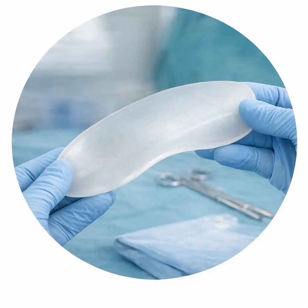
Pectus Implant
A custom-designed silicone implant, shaped precisely to the patient’s chest from a CT scan, is placed beneath the pectoral muscles to fill the depression. It is a cosmetic-corrective rather than structural repair — the sternum itself is not repositioned. A smaller and less invasive procedure than corrective surgery, it is particularly well suited to patients whose primary goal is cosmetic improvement, and is also used in selected revision cases.
Verified patient recommendations
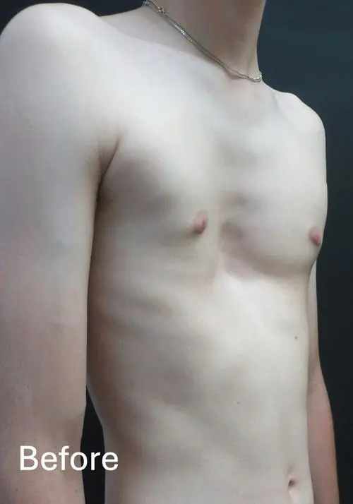
All patients shown have given consent for educational use. Results depend on age, deformity type, severity and treatment chosen.
“I am completely happy with my pectus repair. Great job!”
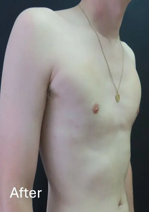
“Took a while to get used to the feeling of the vacuum bell to start, but glad I kept going with it. Very happy with how my chest has improved in appearance so far. I feel more confident in my appearance now.”
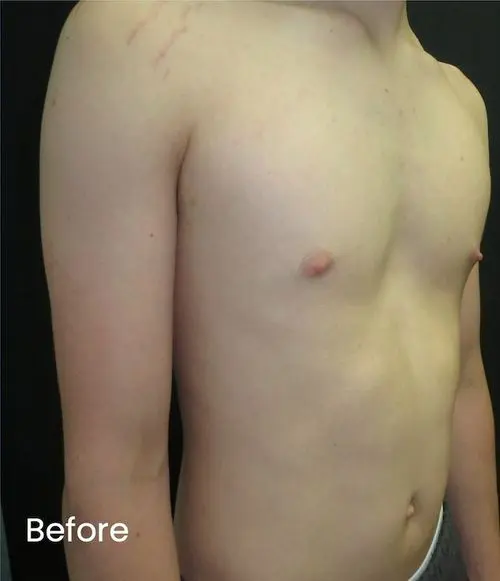
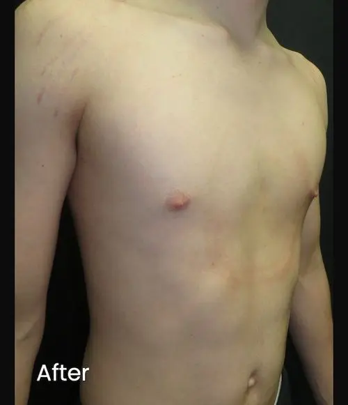
“We're very pleased with the results and the amazing support we've had from Mr Hunt.”
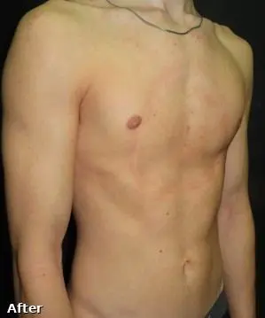
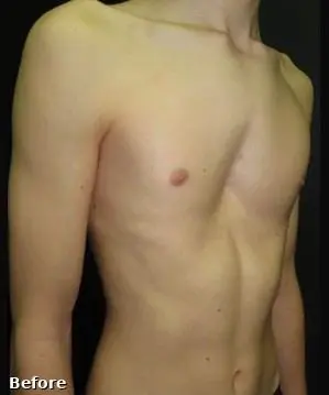
“Much improvement after years of physical difficulties. The treatment gave me many improvements after just 6 months.”
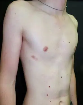
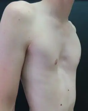
“The experience has been brilliant. Since the surgery I have noticed improvements in my fitness and am happier about how it looks.”
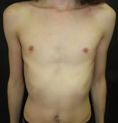
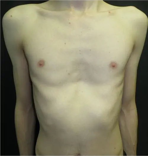
“Feels natural. Part of life. Doesn't hurt. Better than I could imagine.”
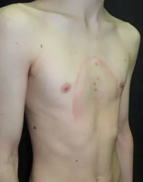
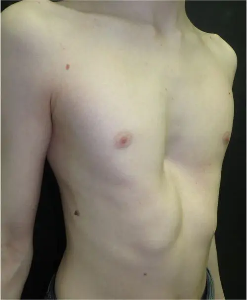
“Mr Hunt has been really supportive and understanding. He has explained every step and reassured me along the way. I am so pleased with the result of surgery and feel like a new man. I can now live my life feeling confident in my own skin.”
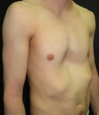
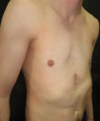
“Excellent procedure for severe, asymptomatic pectus excavatum. I have gained much more confidence in my body and self-image and would highly recommend pectus implant.”
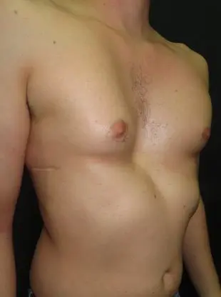
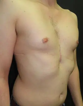
“Since having the implant surgery, I honestly feel like I've got a new lease of life. I can wear clothes I'd never have worn before, take my top off without thinking twice, and generally just feel so much more confident in myself. I honestly can't thank Mr Hunt enough.”
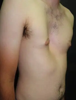
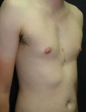
“I wear my Bell overnight and now don't notice. It now feels weird to sleep without. Very happy with the result. This summer I have been swimming with my friends and they didn't notice anything. I'm no longer considering surgery and I am happy with my chest.”
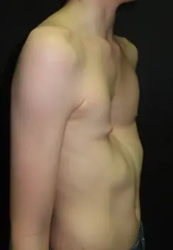
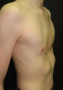
“It has improved my rib cage sticking out massively and it hasn't been that painful.”
Concerned about pectus excavatum?
Every assessment begins with careful examination, clear explanation and honest discussion about the options. If treatment isn't indicated, Mr Hunt will say so. If it is, you will understand exactly why, what it involves and what to expect.