
Mixed & Asymmetrical Pectus
Combined deformity
Chest wall deformities combining features of pectus excavatum and pectus carinatum — often in an asymmetric pattern. Specialist assessment and individualised surgical planning are central to managing these complex presentations.
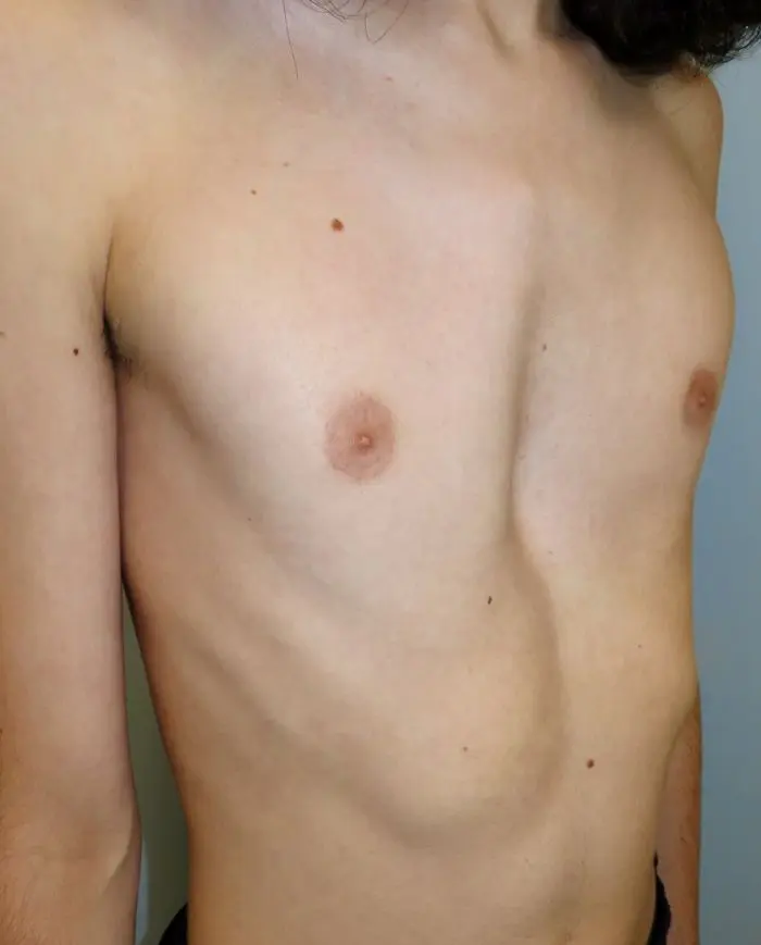
“ Mixed and asymmetrical pectus can look quite different between patients, but the common thread is a chest wall that needs careful three-dimensional analysis before any treatment is planned. The deformity is often more complex than it first appears, and the operative strategy has to be built around the individual anatomy. ”
Mr Ian Hunt — Consultant Thoracic & Chest Wall Surgeon
What is mixed & asymmetrical pectus?
Mixed pectus describes a chest wall deformity in which features of pectus excavatum and pectus carinatum coexist — typically in an asymmetric pattern that does not conform to either condition alone.
One side of the chest may be depressed while the other is prominent. The sternum may be rotated or tilted within the thoracic cage rather than simply pushed inward or forward. In some patients, the central sternal contour is relatively neutral but there is marked lateral asymmetry, with differing costal cartilage morphology on each side. The overall appearance is of an irregular, asymmetric chest wall that is immediately noticeable to the patient but may be difficult to classify.
Because the presentation does not conform to a standard pectus pattern, scoring indices such as the Haller index — designed for isolated pectus excavatum — are often less informative. Characterising the anatomy requires thorough clinical examination and, in most surgical candidates, CT imaging with 3D reconstruction. Treatment planning is correspondingly more involved, and surgical correction is technically more demanding than for isolated PE or PC.
At a glance
Mixed and asymmetrical pectus is not a single named entity but a descriptive category. The underlying anatomy varies considerably between patients. A careful individual assessment — rather than application of a standard protocol — is the starting point for any treatment discussion.
The anatomy of mixed & asymmetrical pectus
There is no single anatomical pattern — mixed and asymmetrical pectus describes a spectrum. What links these presentations is that the chest wall does not conform to isolated PE or PC, and the asymmetry between the two sides is a consistent and clinically important feature.
A spectrum of combined deformity
In the most common pattern, the sternum is rotated within the thoracic cage — tilted so that one hemithorax appears depressed while the other appears relatively prominent. The costal cartilages may differ markedly in length and angulation between left and right, contributing to a visible and palpable asymmetry that often extends beyond the sternum itself to involve the lateral chest wall and lower rib cage.
In other presentations, there is a true combination of excavatum and carinatum features — a central lower sternal depression co-existing with bilateral or unilateral cartilage overgrowth producing some degree of protrusion. The two components may be of similar severity, or one may clearly predominate. Rib flare is a common co-existing finding that further alters the apparent chest wall contour.
Because no single index adequately captures this range of anatomy, the 3D CT remains the most informative investigation. It defines sternal rotation, quantifies intrathoracic encroachment, and maps the individual costal cartilage morphology on each side — all of which inform operative planning in ways that clinical examination and standard CT slices alone cannot.
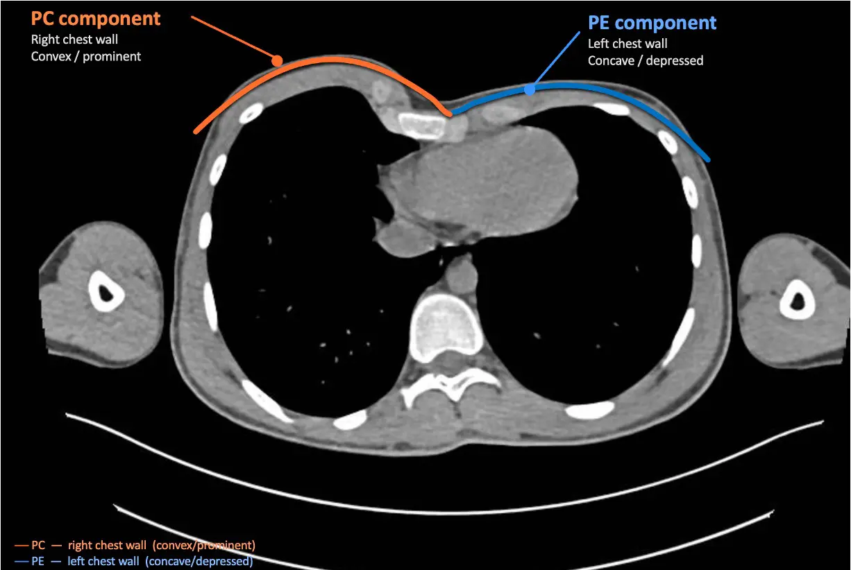
Axial CT chest demonstrating mixed pectus deformity — PC component right side (orange, convex/prominent) and PE component left side (blue, concave/depressed). Reproduced with patient consent.
Why asymmetry matters
Asymmetric deformity introduces surgical considerations that do not arise with symmetric PE or PC. Correcting one component in isolation — for example, addressing only the depressed side — risks worsening the overall contour or creating new asymmetry on the opposite side.
Operative planning for asymmetric pectus typically involves modelling the correction in three dimensions before surgery, with strategy agreed jointly with the patient. Where the primary concern is appearance rather than function, and the degree of structural abnormality is modest, a non-corrective implant may offer a simpler alternative to open chest wall surgery.
In complex or revision cases — for example, where prior surgery at another centre has left residual or recurrent asymmetric deformity — the operative challenge is greater and planning more involved. These cases are discussed within a specialist multidisciplinary framework.
The sternum may be rotated, tilted, or angulated — creating asymmetry across the midline rather than simple depression or protrusion
Costal cartilage length and angulation differ between left and right — overgrowth on one side produces the carinatum component; relative underdevelopment the excavatum
Rib flare, chondral asymmetry, and lateral chest wall irregularity frequently accompany the sternal abnormality and contribute to the overall appearance
No single measurement (such as the Haller index) adequately captures asymmetric or combined anatomy — 3D CT reconstruction is the essential planning tool
Signs and symptoms
Mixed and asymmetrical pectus presents differently between patients and can be difficult to recognise without specialist assessment. The asymmetric element is often more distressing to patients than the absolute degree of depression or protrusion.
Chest wall appearance & psychological impact
The visible asymmetry is typically the most distressing aspect of the presentation. The chest may appear to have a depression on one side and a prominence on the other, or to be visibly rotated when viewed from the front. Some patients have lived with the asymmetry for many years before seeking a specialist opinion, often having been told that the appearance is normal variation or would correct with growth. The psychological burden — particularly in adolescents — is frequently significant.
Functional symptoms
Breathlessness on exertion, reduced exercise tolerance, and chest tightness are reported in a proportion of patients — particularly where the excavatum component is significant and encroaches on intrathoracic structures. Musculoskeletal chest discomfort, often related to asymmetric costal cartilage loading or rib flare, is also common. Symptoms vary considerably in severity and are not always proportional to the apparent degree of deformity.
Associated conditions
Mixed and asymmetrical pectus may be associated with rib flare, scoliosis, Marfan or Ehlers–Danlos syndrome, or Poland syndrome. Where connective tissue features are present, a cardiac screen including echocardiography is arranged as part of the assessment. Some patients have had prior chest wall surgery — including Nuss bar insertion — elsewhere, and present with residual or recurrent asymmetric deformity. These revision cases require particularly careful assessment.
Diagnostic delay is common in mixed pectus. Because the presentation does not fit either standard pectus type clearly, patients are sometimes reassured that the chest is normal or that the deformity will self-correct. Where an asymmetric chest wall causes symptoms or distress, specialist review is appropriate regardless of whether a previous opinion was sought.
How mixed pectus presents
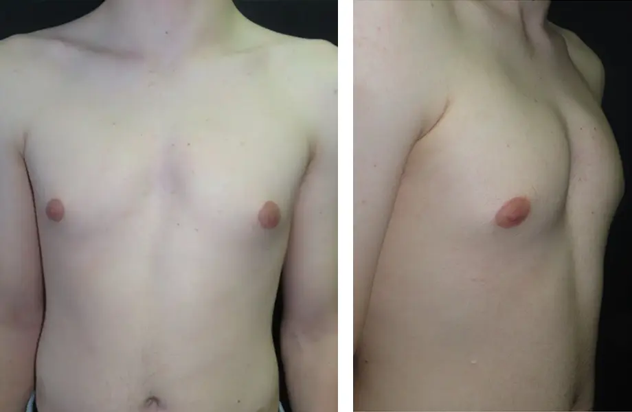
The appearance of mixed and asymmetrical pectus varies considerably between patients. Because the deformity does not conform to a single pattern, it is often difficult to assess without clinical examination and imaging. The examples below illustrate a range of presentations encountered in specialist practice. All photographs are reproduced with full written patient consent.
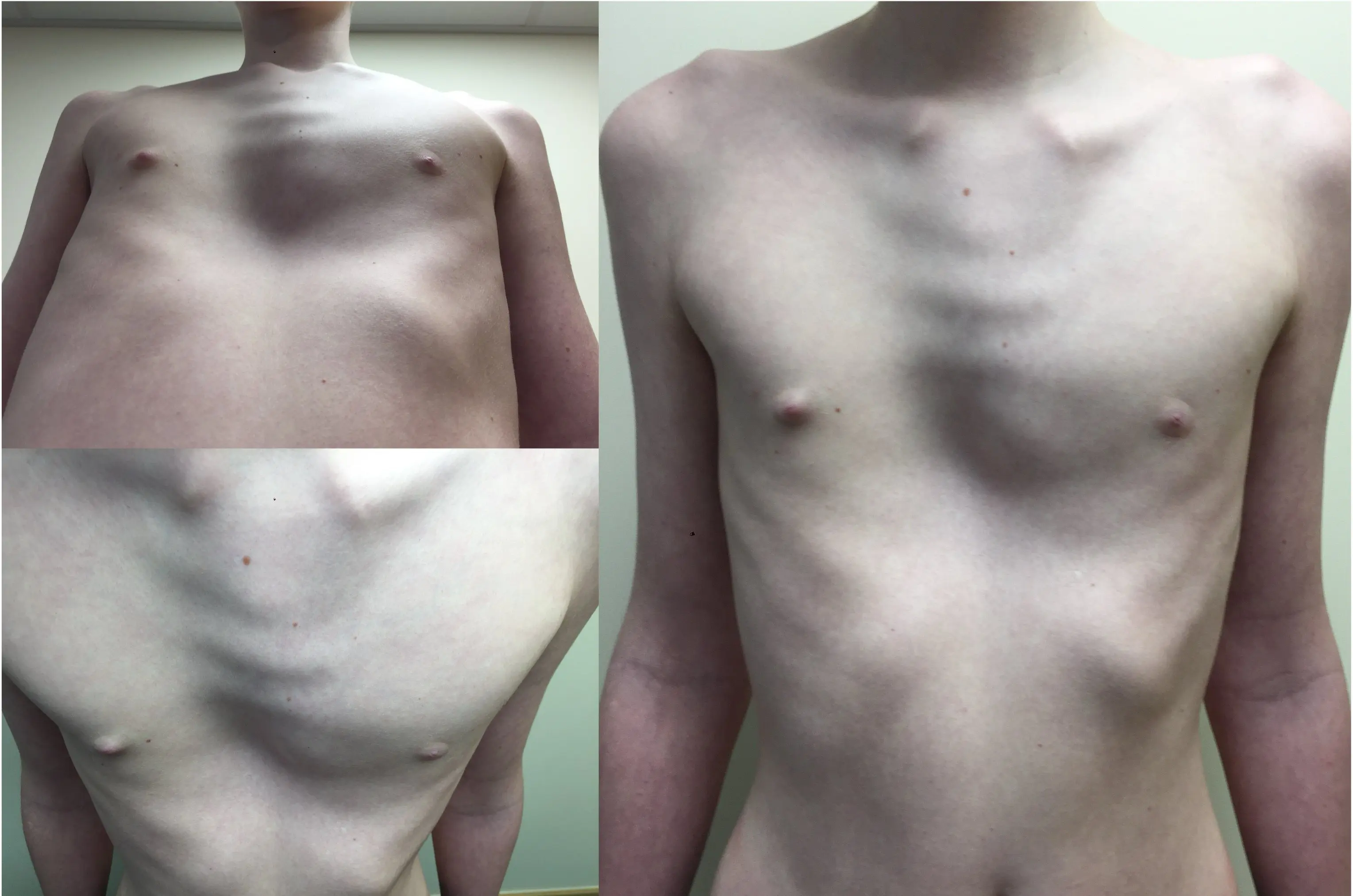
Mixed deformity — PE-dominant, mild–moderate
Three views demonstrating a mild-to-moderate mixed pectus with a predominant excavatum component. Frontal views show lower sternal depression with asymmetric rib cage contour; the oblique view highlights sternal rotation and relative prominence on the opposite side. The combined deformity is visually apparent and often causes significant distress, but may not have been formally recognised at routine review.
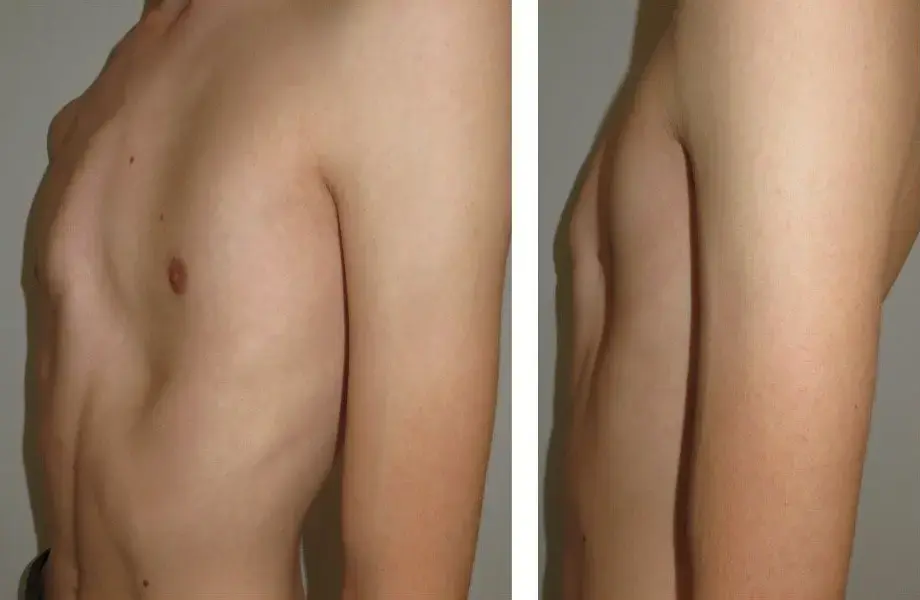
Mixed deformity — PC-dominant, mild–moderate
Two lateral views demonstrating a mild-to-moderate mixed pectus with a predominant carinatum component. The lateral profile shows anterior chest wall protrusion with underlying asymmetry — the prominence is not uniformly distributed, and sternal tilt is apparent on closer inspection. Management follows the carinatum pathway, with the asymmetric element factored into bracing or surgical planning.
Bilateral carinatum — horseshoe pattern
Frontal and oblique views showing moderate bilateral carinatum in a horseshoe pattern. The bilateral protrusion creates an apparent central concavity that can, at first glance, suggest a mixed or excavatum component — but on examination and imaging the sternum is not depressed. Assessment and management follow the carinatum pathway, accounting for the bilateral and symmetric nature of the deformity.
How it is assessed
Mixed pectus requires more careful assessment than isolated pectus excavatum or pectus carinatum. Because the deformity does not conform to a single standard pattern, the first clinical step is identifying which component is dominant — and that finding shapes the entire direction of assessment and treatment.
Identifying the dominant component
Most patients with mixed pectus have one component that is structurally and clinically more significant than the other. Where the predominant feature is sternal depression, the deformity is classified as excavatum-dominant. Where the predominant feature is sternal or cartilage protrusion, it is classified as carinatum-dominant. Once the dominant type is identified, assessment follows the same pathway used for that condition — with the secondary component characterised alongside. Patients can generally be offered the same treatment options as those with isolated PE or PC, though planning is more involved.
Assessed as pectus excavatum
When the dominant feature is sternal depression, assessment follows the same structured pathway used for pectus excavatum. Asymmetry is formally documented and its implications for operative planning are addressed as part of that assessment.
- Full clinical examination, photographic documentation in standardised positions
- CT chest with 3D reconstruction — Haller index and correction index measured, though these may underrepresent severity when significant asymmetry is present
- Echocardiogram and pulmonary function testing where clinically indicated
- Cardiology and respiratory review for selected patients with functional symptoms
- Dedicated surgical consultation with three-dimensional review of CT anatomy
Assessed as pectus carinatum
When the dominant feature is sternal or cartilage protrusion, assessment follows the pectus carinatum pathway. Skeletally immature patients are assessed for bracing candidacy; older patients or those with more complex anatomy are assessed with a view to surgical correction where indicated.
- Full clinical examination, photographic documentation
- Chest X-ray and/or CT imaging depending on age, severity, and treatment goals
- Bracing assessment for growing patients — severity, brace tolerance, and compliance discussed
- Spirometry and functional assessment where relevant
- Surgical consultation for patients not suitable for, or not wishing to proceed with, bracing
Additional assessment considerations for mixed deformity
The following considerations are relevant in most mixed and asymmetrical presentations, and are discussed at the initial consultation.
3D CT reconstruction — particularly where surgery is being considered
Standard indices such as the Haller index are designed for symmetric sternal depression and may underrepresent the severity of a mixed or asymmetric deformity. Where surgery is being considered, 3D CT reconstruction is the most useful investigation — it defines sternal rotation, maps costal cartilage morphology on each side, and provides the anatomical detail that operative planning requires. For patients managed non-surgically, CT may not be needed, and a clinical assessment with or without plain imaging is often sufficient.
The secondary component is assessed alongside the dominant one
Even in a clearly excavatum-dominant or carinatum-dominant presentation, the non-dominant component is characterised as part of the overall assessment. Its degree of depression, protrusion or asymmetry is relevant to operative strategy — influencing decisions about bar placement, costal cartilage resection, and whether implant reconstruction may be needed to address the overall contour.
Operative planning tends to be more involved than for isolated conditions
Mixed and asymmetrical deformity is generally more complex to correct than isolated PE or PC. Where surgery is indicated, the operative strategy is built around the three-dimensional CT anatomy and agreed with the patient before proceeding. It will often differ substantially from what would be planned for a symmetric presentation of either type, and takes longer to define.
In some patients no single component clearly dominates, and the deformity is genuinely balanced or symmetrically mixed. These cases do not fit neatly into either standard pathway and are assessed entirely on their individual anatomy, symptoms, and priorities. Operative planning is based on full 3D CT review and may involve a bespoke surgical strategy that draws elements from both PE and PC correction techniques.
Treatment options
Treatment is tailored to the individual. The decision depends on the severity and nature of the combined deformity, functional symptoms, psychological impact, and the patient’s own goals. Not every patient requires surgery, and not every deformity is best addressed by open correction.
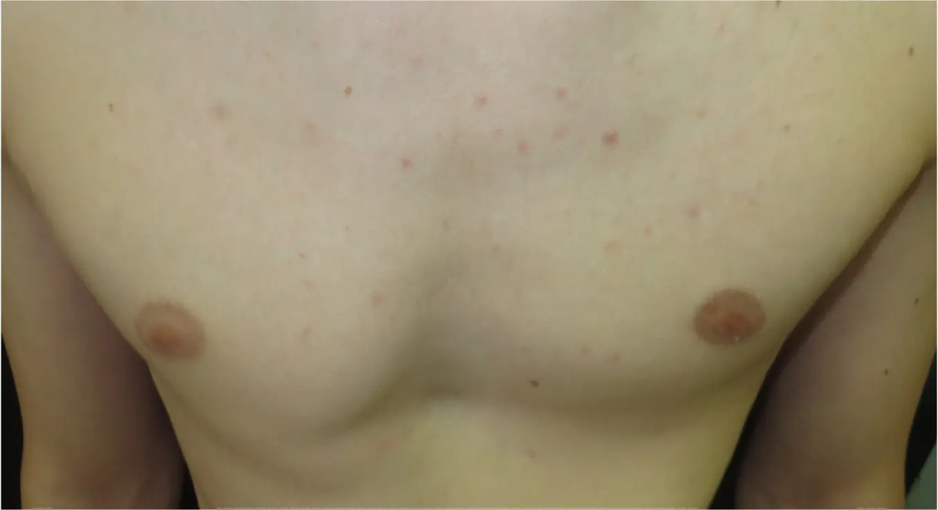
Observation & monitoring
For patients with mild deformity and no significant functional symptoms, active monitoring is often the appropriate initial approach — particularly in adolescents who are still growing. Periodic clinical review documents any change in the deformity and allows the need for intervention to be reassessed as the chest wall matures.
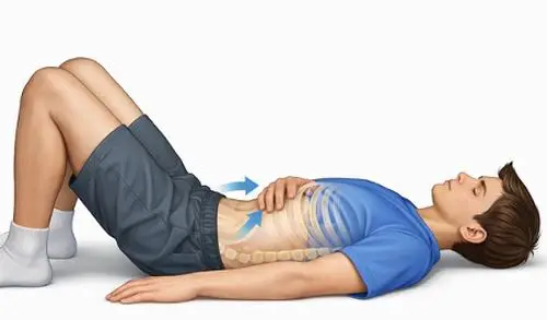
Targeted exercise & postural retraining
A structured physiotherapy programme focusing on core strengthening, postural alignment, and chest wall muscle activation. It will not correct the underlying deformity but can reduce musculoskeletal discomfort and improve functional capacity — both as a standalone measure and as preparation for or recovery from surgery.
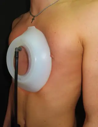
Vacuum bell therapy
Where the deformity is PE-dominant, vacuum bell therapy may have a role in selected skeletally immature patients. The device applies a controlled suction force to lift the anterior chest wall, used daily over an extended period. Results are most reliable in younger patients with flexible chest walls; the asymmetric element may limit the degree of correction achievable.
External bracing
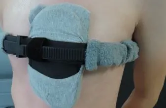
Where the deformity is PC-dominant, external bracing follows the same principles as for isolated pectus carinatum. A custom-fitted brace applies graduated pressure to the prominent cartilage and is most effective in growing patients who can wear it consistently. The asymmetric element is factored into brace design and fitting, with regular review and adjustment.
Surgery is considered when deformity is causing significant symptoms or psychological distress, when non-surgical measures are insufficient, or when the anatomy is clearly best addressed by structural correction. The approach is guided by the dominant type and the individual anatomy. All options are discussed in detail at the surgical consultation.
PE-dominant approaches
Nuss procedure (MIRPE)
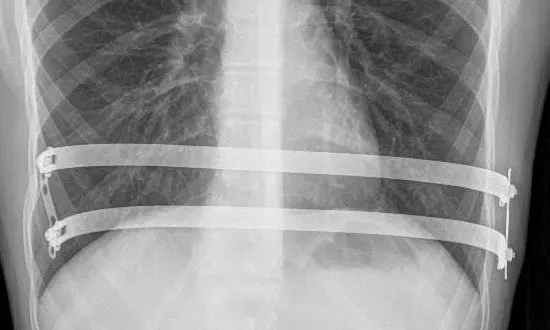
Where the deformity is PE-dominant with a significant sternal depression, the Nuss procedure — minimally invasive placement of a retrosternal support bar — follows the same principles as for isolated pectus excavatum. The bar is placed under thoracoscopic guidance and removed after two to three years once the chest wall has remodelled. In mixed deformity the bar position and curvature may be adjusted to account for the asymmetric component.
Modified Ravitch procedure
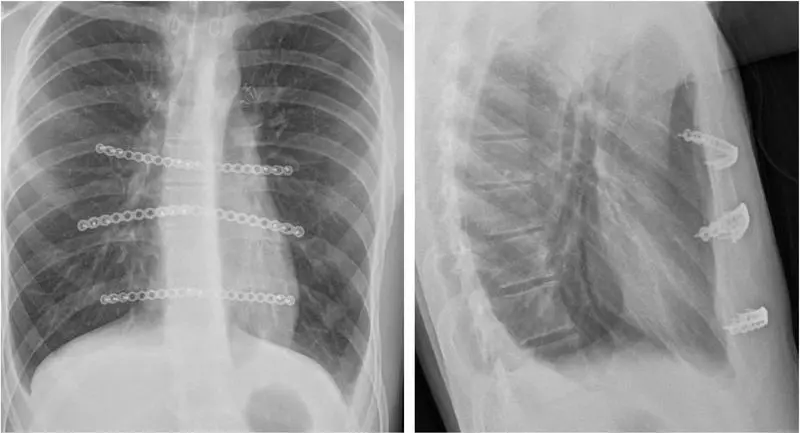
Open chest wall reconstruction involving resection of the abnormal costal cartilages with sternal repositioning. In PE-dominant mixed pectus, the Ravitch approach allows the surgeon to address asymmetric cartilage anatomy directly — correcting both the depressed and any prominent components at a single operation. It offers greater flexibility than bar insertion alone where the anatomy is complex or asymmetric.
PC-dominant approaches
Costal cartilage resection
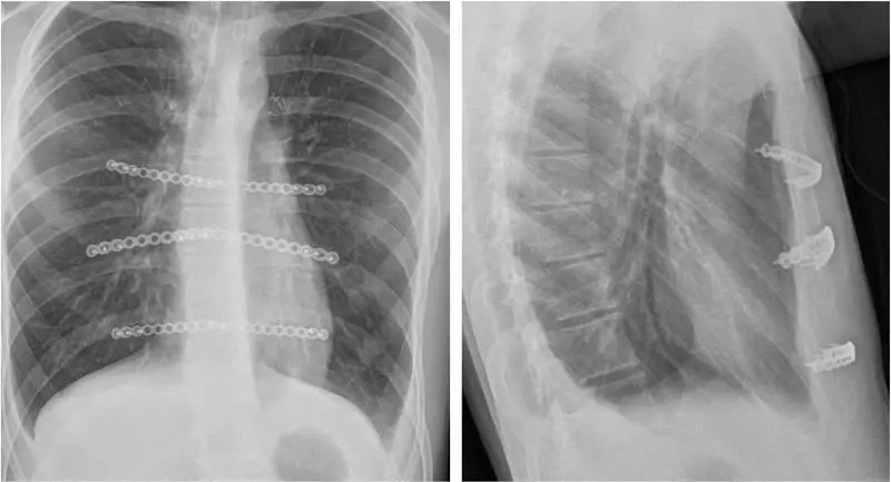
For PC-dominant mixed pectus, surgical correction centres on resection of the overgrown costal cartilages responsible for the anterior protrusion. The approach is similar to the carinatum component of a modified Ravitch procedure, tailored to the individual anatomy. Where asymmetry is present, the cartilage resection is planned to account for the combined deformity, avoiding over-correction on the prominent side or worsening of the depressed component.
All presentations
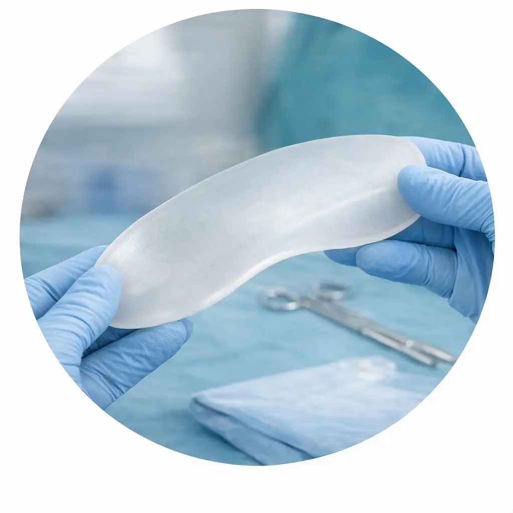
Custom chest wall implant
A patient-specific silicone implant, designed from CT data, can fill the depressed component of a mixed or asymmetric deformity without skeletal correction. This option avoids open chest surgery and the associated recovery, and may suit patients whose concern is primarily aesthetic and where the structural abnormality is modest. Placed through a small incision, typically as a day case or short-stay admission.
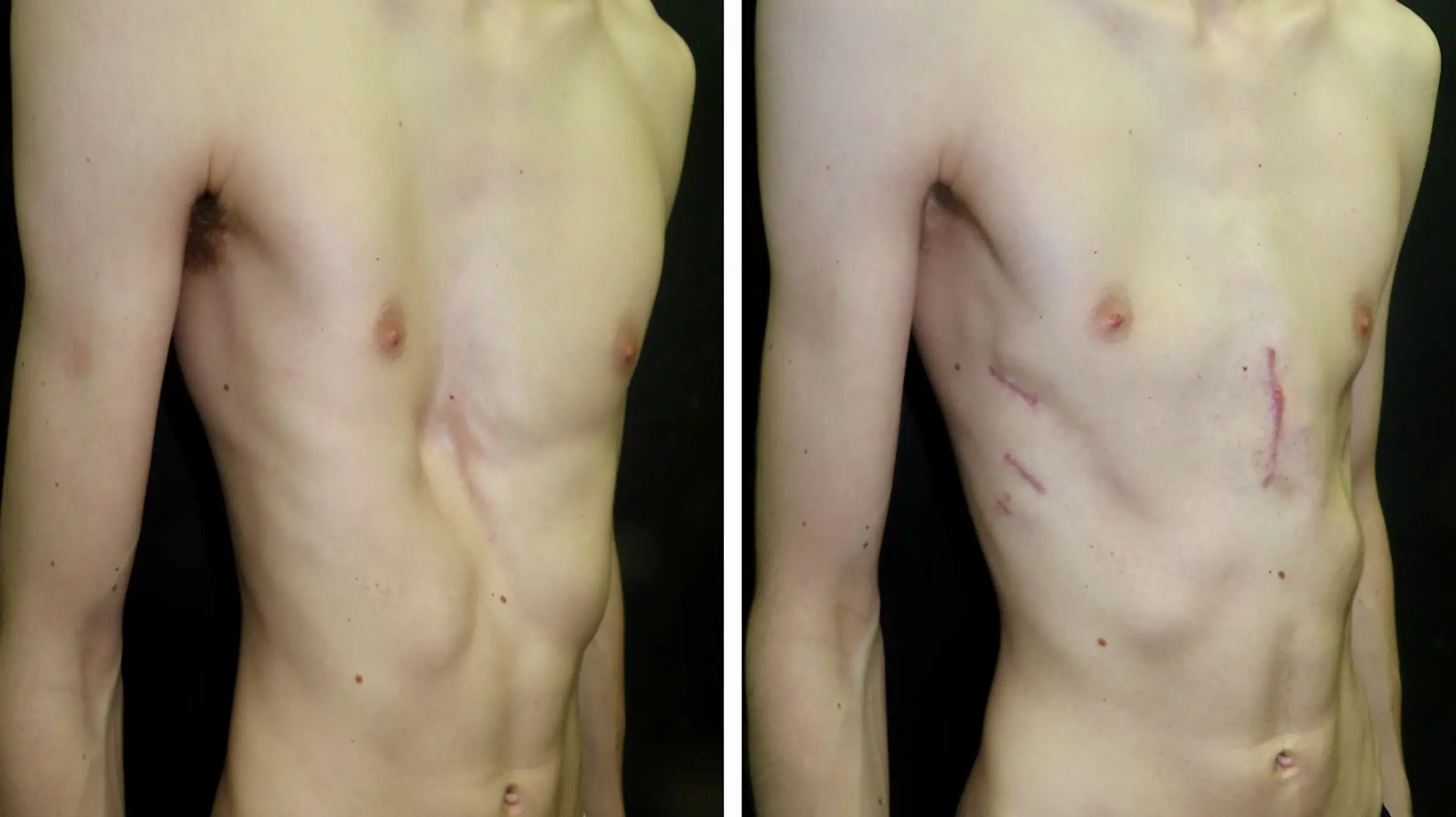
Hybrid reconstruction
A combined approach using limited costal cartilage resection alongside internal bar fixation to address both components of the deformity in a single operation. Hybrid techniques aim to correct the excavatum and carinatum elements while minimising the extent of dissection required compared to a full open reconstruction. Case selection and operative planning are based on detailed CT analysis; not all mixed pectus presentations are suitable.
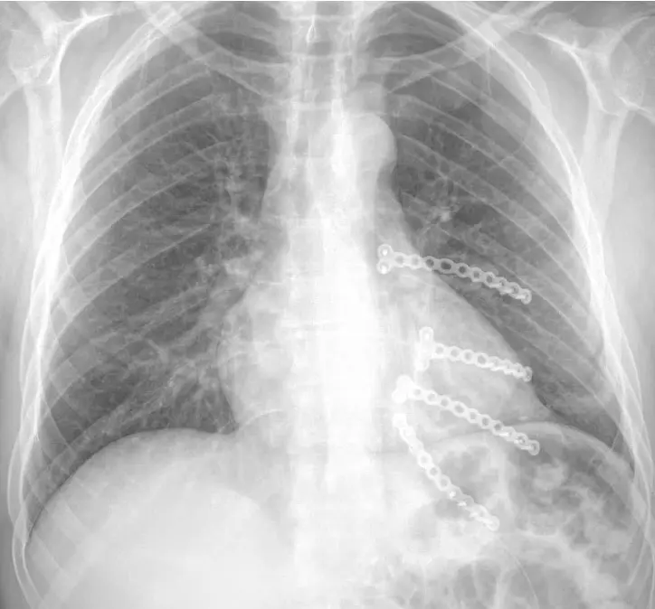
Complex & revision surgery
Some patients with mixed or asymmetric pectus present with prior chest wall surgery — including Nuss bars placed elsewhere — and residual or recurrent deformity. Others have co-existing conditions such as Poland syndrome or significant scoliosis that add complexity. These cases are managed with an individually modified operative strategy, often discussed within a specialist multidisciplinary framework before any surgical planning is finalised.
Frequently asked questions
Is surgery always required for mixed pectus?
No. Many patients are managed with observation, monitoring, and physiotherapy — particularly during adolescence or where functional symptoms are absent. Surgery is discussed when the deformity is symptomatic, causing significant psychological distress, or is clearly progressive. The decision is made jointly with the patient following a full assessment and detailed discussion of the available options.
Can the Nuss procedure correct a mixed deformity?
Yes — the Nuss procedure can be a good option in PE-dominant mixed pectus, but it requires experience and careful planning. The key challenge is ensuring that bar correction of the depressed component does not inadvertently accentuate the prominent side. This can be addressed through different bar bending techniques, allowing the applied forces to be shaped to the individual chest wall contour. In more complex cases, a hybrid approach — combining bar insertion with targeted cartilage work — can address both the depression and the prominence simultaneously, avoiding excessive worsening of the carinatum element. The right approach depends on the specific anatomy, which is why CT assessment and specialist surgical planning are central to the decision.
Does a mixed deformity worsen over time?
It often becomes more apparent during adolescent growth. Once skeletal maturity is reached, the structural anatomy is largely stable, though symptoms and the psychological impact of the appearance can evolve independently of any change in the actual deformity. Regular clinical review during the growth years is helpful in tracking progression and informing the timing of any decision about intervention.
What does 3D CT show that a standard CT does not?
Standard CT slices show the anatomy in cross-section but do not readily convey the three-dimensional shape of the chest wall or the degree of sternal rotation. 3D surface and bony reconstruction allows the asymmetry to be visualised from any angle, the individual costal cartilage morphology on each side to be assessed, and the operative correction to be planned in three dimensions before the patient reaches the operating table. It is an essential part of the pre-operative assessment for mixed pectus.
Is there a role for bracing in mixed pectus?
Yes — bracing can be a good option in PC-dominant mixed pectus, but it works best in experienced hands. The key challenge is applying enough pressure to address the prominent component without inadvertently worsening the depressed side. This requires careful brace design: different bar bending techniques allow the applied forces to be directed precisely, avoiding compression over any area of depression. In more complex presentations, a hybrid approach — combining elements of standard bracing with customised modifications — can address both components simultaneously. As with all treatment decisions in mixed pectus, the choice of brace configuration should follow specialist assessment of the individual deformity pattern.
Can vacuum bell therapy be used in mixed pectus?
It can be considered in PE-dominant cases, but it is technically more challenging than in isolated pectus excavatum. The vacuum bell works by creating negative pressure over the depressed sternum, drawing the chest wall outward over time. In a symmetric PE, the device sits centrally and maintains consistent contact. In mixed or asymmetric pectus, the uneven chest wall contour makes it harder for the vacuum bell to achieve and sustain an effective seal, and the device may shift position with use — reducing both the force applied and the consistency of correction. This does not exclude it as an option, but it does mean that patient selection needs to be careful, and expectations should be realistic. It tends to work best in younger, growing patients with a predominantly central depression and relatively mild asymmetry.
Will my insurance cover treatment?
Coverage depends on the insurer and the clinical indication. Treatment for symptomatic or functionally significant mixed pectus is more likely to attract insurance support than purely cosmetic correction. The clinic’s administrative team can assist with pre-authorisation queries. Further information is available on the Fees & Funding page.
Arrange a specialist assessment
All patients are seen by Mr Ian Hunt personally. Consultations include a full clinical examination, review of any existing imaging and a clear, unhurried discussion of the options available.