
Poland syndrome
A rare congenital condition characterised by underdevelopment or absence of the pectoralis major muscle, with a clinical spectrum that ranges from subtle chest asymmetry to significant chest wall and rib involvement.

A congenital spectrum, not a single diagnosis
Poland syndrome is a rare congenital chest wall condition characterised by underdevelopment or absence of the pectoralis major muscle, with or without associated abnormalities of the chest wall, ribs, soft tissues, breast, nipple and hand. It is always present from birth, and is not acquired or degenerative in origin.
The condition affects males more commonly than females and most commonly involves the right side, though left-sided cases occur. The cause is not fully understood but is thought to relate to disruption of blood supply to the subclavian artery during early fetal development.
In women, Poland syndrome typically produces breast and nipple asymmetry alongside the pectoral deficit, and requires careful consideration of both functional and aesthetic aspects when planning treatment. Female patients represent an important and sometimes overlooked group within this spectrum.
Poland syndrome is best understood as a spectrum. At one end, a patient may have subtle underdevelopment of part of the pectoralis major with almost no visible asymmetry; at the other, there may be complete absence of the pectoralis muscles combined with rib and costal cartilage abnormalities, significant soft-tissue and breast asymmetry, and occasionally hand differences.
Key facts
Mild Poland syndrome
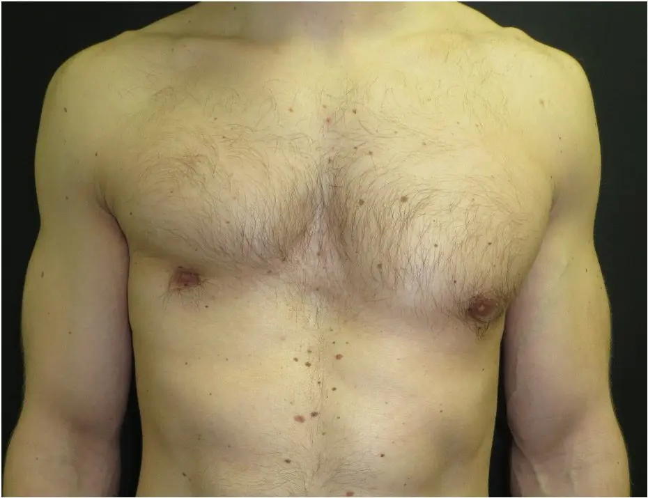
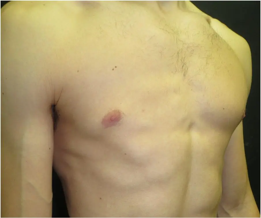
In mild cases, only part of the pectoralis major is underdeveloped — typically the sternocostal head, which forms the lower and inner portion of the muscle. The clavicular head, which runs to the shoulder, is often intact or near-normal.
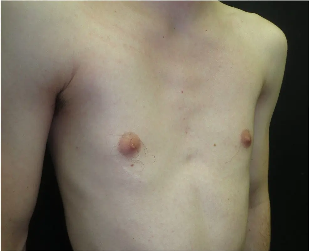
The chest asymmetry in mild Poland syndrome can be subtle and may not become apparent until late childhood or adolescence. In some patients it is only recognised during a routine examination or following a sports injury. Nipple position is usually normal or only mildly displaced. There is no structural involvement of the ribs or costal cartilages.
Patients with mild Poland syndrome are typically asymptomatic from a functional standpoint. Many are aware of the asymmetry and seek assessment for cosmetic or psychological reasons — particularly in adolescence or early adulthood when body awareness increases. In those who wish treatment, a custom soft-tissue implant placed beneath the pectoral fascia can restore natural chest contour without the need for complex reconstruction.
Moderate Poland syndrome
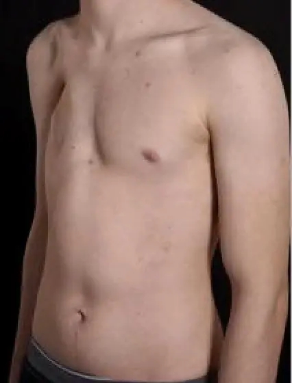
In moderate cases, the pectoralis major is more extensively absent or hypoplastic. The pectoralis minor, serratus anterior and latissimus dorsi may also be underdeveloped, producing a more pronounced hollow or flat appearance to the anterior chest wall.
In females, moderate Poland syndrome produces asymmetric breast development alongside the pectoral deficit. The breast on the affected side is typically significantly smaller, and the nipple and areola may be higher, smaller or laterally displaced. This combination of pectoral and breast asymmetry is the defining feature of Poland syndrome in women and is an important factor in treatment planning.
In males, the pectoral region lacks the normal muscular contour, most apparent when the arms are raised or when viewed from directly in front. The ribs and costal cartilages are structurally normal in moderate cases. Assessment includes CT imaging and 3D surface scanning to precisely define the soft-tissue and muscle deficit before designing a custom implant.

Severe Poland syndrome
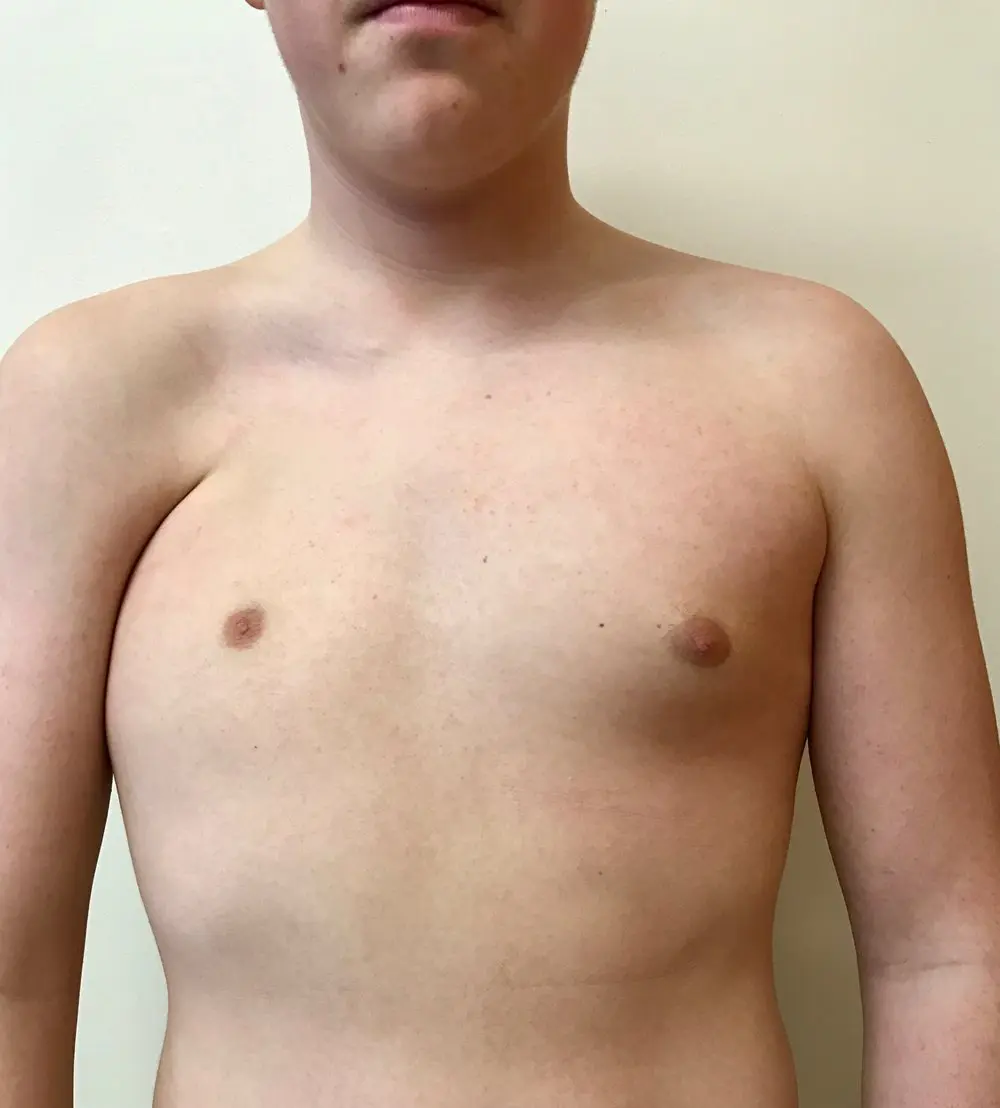
In the most severe cases, Poland syndrome involves not only complete absence of the pectoralis major and associated soft tissues but also structural abnormalities of the ribs and costal cartilages on the affected side.
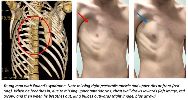
The ribs may be hypoplastic, fused or partially absent. When the upper anterior ribs and costal cartilages are missing, the chest wall becomes structurally deficient — causing paradoxical inward movement during inspiration and outward bulging during expiration. In females there may be complete absence of breast tissue on the affected side.
In a proportion of patients with severe Poland syndrome, hand differences are also present — most commonly brachydactyly or syndactyly on the affected side. Shoulder hypermobility and inferior displacement may also occur where the pectoral clavicular attachment is absent.
CT imaging with 3D reconstruction is essential to characterise the bony anatomy before surgical planning. Reconstruction is typically staged — rib and chest wall repair followed by soft-tissue or breast reconstruction.

Severe Poland syndrome — rib-absent chest wall
The illustration and clips below are specific to rib-absent Poland syndrome. The clinical illustration shows the structural deficit; the video demonstrates paradoxical chest wall movement during breathing; the CT sequence maps the missing skeletal structures essential for surgical planning.
Poland syndrome in women
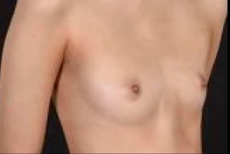
In female patients, Poland syndrome most commonly presents as breast and nipple asymmetry on the affected side. The pectoralis major is absent or hypoplastic, and breast development — which occurs above the pectoral muscle — is directly affected, producing asymmetry that often becomes apparent during adolescence.
The spectrum ranges from subtle breast volume difference and mild nipple displacement to complete unilateral breast absence. When Poland syndrome coexists with a pectus deformity, the combined chest wall and soft-tissue asymmetry is more complex. CT imaging with 3D reconstruction is required to characterise both components before reconstruction is planned.
Surgical options for female Poland syndrome include custom soft-tissue implant, fat grafting, or staged reconstruction depending on the degree of breast and chest wall involvement. Assessment of the skeletal anatomy is essential before any breast reconstruction to ensure the correct foundation.
Poland-like & acquired pectoral deficiency
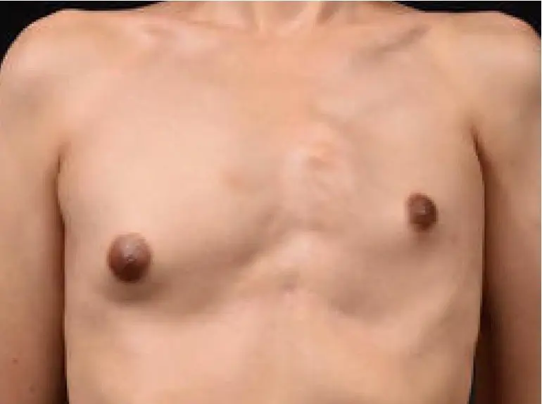
Not all pectoral asymmetry is true Poland syndrome. This category covers two distinct groups: congenital pectoral underdevelopment that resembles Poland syndrome but lacks its defining features, and acquired pectoral deficiency caused by injury, surgery, or progressive muscle degeneration.
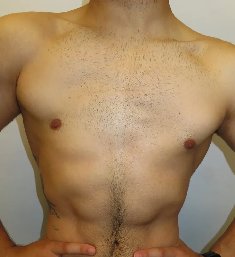
In congenital pectoral underdevelopment, the pectoralis major is hypoplastic or partially absent from birth, but there is no rib or costal cartilage involvement, no hand difference, and the pattern of muscle loss does not follow the typical Poland distribution. These patients often present in the same way as mild Poland syndrome and require the same structured assessment before any treatment is planned.
Acquired pectoral deficiency develops after birth and has a clear precipitating cause: pectoralis rupture following heavy loading (bench press injury), progressive degenerative muscle atrophy, post-surgical resection, or radiotherapy-related soft-tissue loss. The chest wall skeleton is structurally normal and hand differences are absent.
Distinguishing these categories matters because treatment planning differs. In acquired degenerative cases, the underlying cause must be addressed first. Reconstruction options — fat grafting, custom implant, or soft-tissue flap — are planned individually following full imaging and clinical assessment.
Custom Poland implant reconstruction
For patients with mild to moderate Poland syndrome, a custom-designed chest wall implant is the most reliable means of restoring chest wall contour and symmetry. The implant is individually manufactured to match the precise shape and volume deficit of each patient.
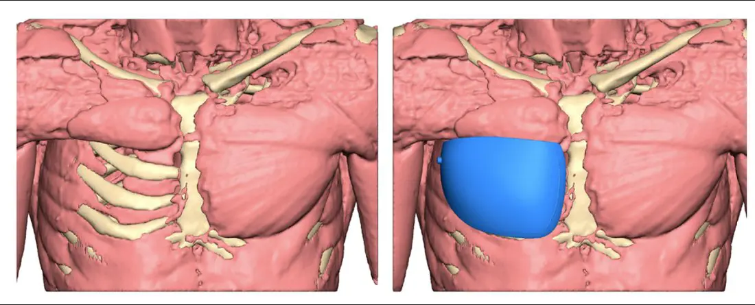
The implant is planned from the patient's own CT scan using 3D modelling software. The deficit is mapped precisely, and a silicone implant is designed to fill the exact volume and surface contour deficiency — including any asymmetry in the pectoral border, infraclavicular hollow or nipple level. The result integrates naturally with the existing anatomy without muscle transfer or large incisions.
The procedure is performed under general anaesthesia through a small axillary incision, typically as a day case or overnight admission. Recovery is straightforward, with most patients returning to desk work within one to two weeks and full activity by six to eight weeks.
- 1CT imaging & 3D planningThin-slice CT chest provides anatomical data for implant design. Volume deficit and surface contour are mapped in three dimensions.
- 2Implant design & approvalA custom silicone implant is designed and reviewed with the patient before manufacture. The 3D model is signed off before fabrication.
- 3Surgical placementThe implant is placed through a small axillary incision under general anaesthesia, positioned beneath the skin and pectoral fascia.
- 4RecoveryDesk work within one to two weeks. Full activity including sport and gym by six to eight weeks. No long-term restrictions.
Before & after — custom Poland implant
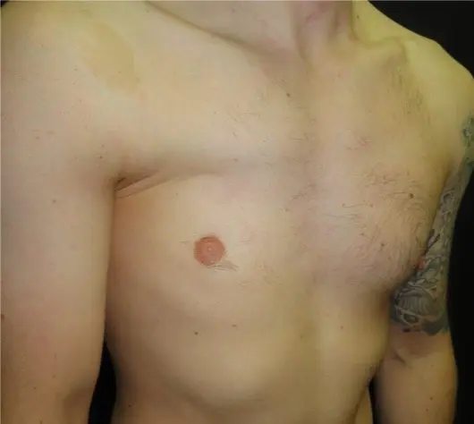
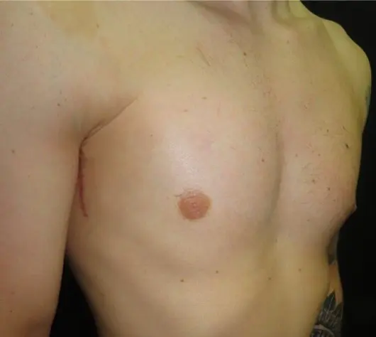
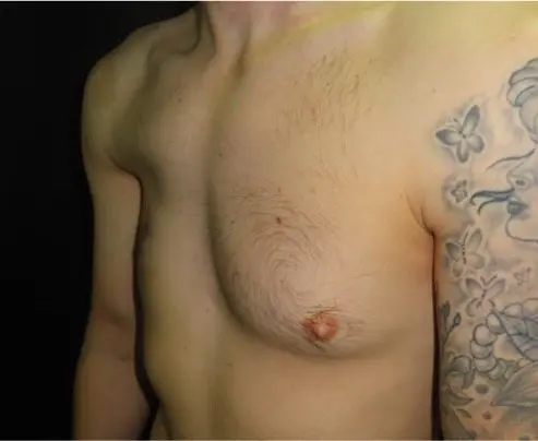
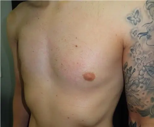
Custom 3D-designed silicone implants. Images reproduced with patient consent.
Structured assessment pathway
Every patient is assessed individually. The consultation begins with a full history — including the age of first noticing the asymmetry, any functional symptoms, family history and personal treatment goals.
Clinical examination is followed by 3D surface scanning and targeted imaging — typically CT chest with 3D reconstruction — to characterise the extent of muscle, soft-tissue and rib involvement. In patients with suspected rib or costal cartilage abnormalities, thin-slice CT allows precise anatomical assessment before planning any reconstruction.
Treatment options are discussed at a dedicated follow-up appointment once imaging is available. Self-referral is accepted. Patients already under the care of another specialist are welcome to seek a second opinion.
Assessment includes
Specialist assessment for Poland syndrome
Every patient is assessed individually, with treatment planning tailored to the degree of chest wall asymmetry, soft-tissue involvement and personal goals. Self-referral is accepted.