
Pectus Arcuatum
Currarino–Silverman syndrome
A rare congenital chest wall deformity involving abnormal angulation of the upper sternum and manubriosternal junction — producing a distinctive curved, angular or wave-like profile. Often misdiagnosed as a mixed pectus deformity, it merits careful specialist assessment and imaging.

“ Not every case of pectus arcuatum requires surgery — but because the deformity combines two opposing sternal abnormalities, the assessment needs to be thorough. Understanding the three-dimensional anatomy and any functional effect is essential before recommending a treatment pathway. ”
Mr Ian Hunt — Consultant Thoracic & Chest Wall Surgeon
What is pectus arcuatum?
Pectus arcuatum is a rare congenital chest wall deformity involving abnormal angulation and shape of the upper sternum and manubriosternal junction. The sternum is typically short and broad, and may have an unusual curved or angular configuration.
The chest may show a prominent upper sternum with a lower inward-sloping component, giving the side profile a distinctive curved, angular or wave-like appearance. Because it can look like a combination of pectus carinatum and pectus excavatum, pectus arcuatum is often misdiagnosed as a mixed pectus deformity. The underlying anatomy — an abnormal upper sternal shape rather than a straightforward combination of two standard deformities — distinguishes it from both.
Careful specialist assessment and imaging are important to define the sternal shape, costal cartilage anatomy, and three-dimensional thoracic structure before making any treatment recommendation. Because the condition is genuinely rare, it is not uncommon for patients to have had their chest wall examined previously without a clear diagnosis being reached.
At a glance
Currarino–Silverman syndrome is the eponymous term sometimes used for pectus arcuatum, particularly in the older literature. The condition was described by Currarino and Silverman in 1958 as a distinct deformity characterised by premature fusion of the sternal segments and superior costal cartilages, producing the combined protrusion-depression pattern.
The anatomy of pectus arcuatum
Pectus arcuatum involves a distinctive structural abnormality of the sternum and upper costal cartilages. Understanding the underlying anatomy is central to planning accurate assessment and appropriate treatment.
An abnormal sternum
The defining feature of pectus arcuatum is an abnormally shaped sternum. The sternum is typically short and broad, with an unusual angular or curved configuration at the manubriosternal junction — the joint between the upper (manubrium) and lower (sternal body) segments.
In most individuals, the sternum runs in a relatively straight line from the clavicles to the xiphoid process. In pectus arcuatum, the manubrium and upper sternal body angle forward, creating a prominent upper chest protrusion. The lower sternal body then curves inward, producing the characteristic lower depression. The net effect is a wave-like or Z-shaped sternal profile when viewed from the side.
The costal cartilages — the cartilaginous segments connecting the ribs to the sternum — are often affected by this abnormal sternal orientation. In some cases, premature fusion of sternal segments and adjacent costal cartilage contributes to the rigid angular configuration. This is why the deformity does not respond to bracing in the same way that pectus carinatum does.

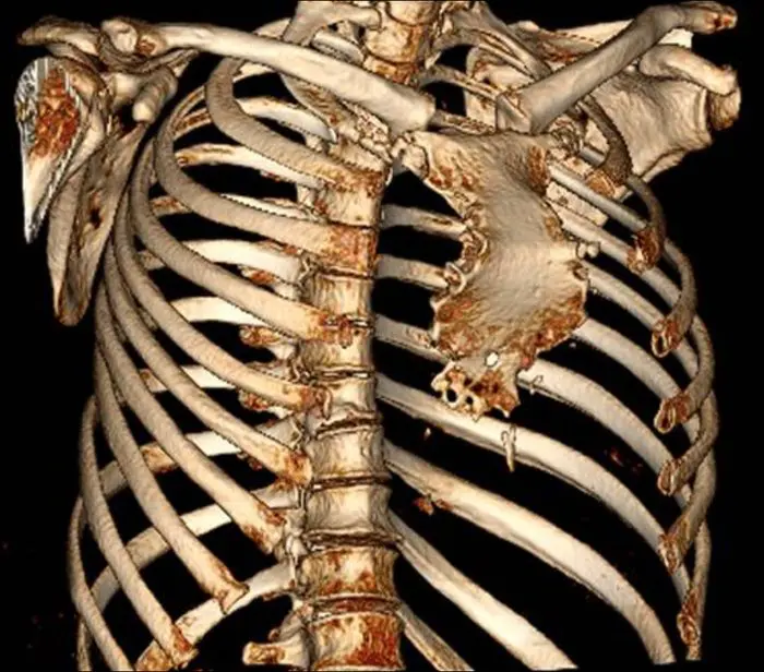
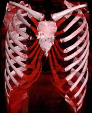
3D CT reconstruction. Anonymised. Reproduced with consent.
Manubrium and upper sternal body angle forward — creating the visible upper chest protrusion
Abnormal angulation at this joint is the structural origin of the wave deformity — often with fusion or premature calcification
Lower sternal body curves posteriorly — producing the inward depression and reduced anterior thoracic volume
Upper cartilages often show abnormal morphology and may be fused, calcified, or maldirected, contributing to rigidity
Signs and symptoms
Pectus arcuatum presents differently from person to person. Some individuals have minimal symptoms; others experience significant functional or psychological difficulty. The combined sternal deformity can affect both the anterior chest wall and the thoracic volume.
Chest wall appearance & psychological impact
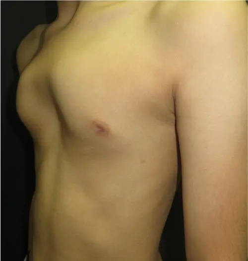
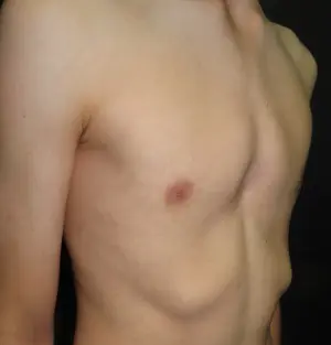
The most consistent finding is the visible wave-like sternal contour: an upper protrusion beneath the upper chest, and a lower depression giving a scooped appearance to the lower sternum. Concerns about chest appearance are often a significant part of the presentation — particularly in younger patients. Difficulty wearing fitted clothing, self-consciousness at swimming or sport, and avoidance of situations that expose the chest are commonly reported.
Chest discomfort
Some patients describe musculoskeletal chest pain, particularly in the costal cartilage region. In cases where rib flare co-exists, the lateral lower ribs may also contribute to discomfort, especially during exercise, prolonged sitting, or bending forwards. Exertional breathlessness and reduced exercise tolerance are also reported when the lower sternal depression reduces intrathoracic volume.
Associated deformities
Pectus arcuatum may occur alongside rib flare, mild scoliosis, or features of connective tissue laxity. A minority of cases have a degree of pectus excavatum in the lower sternal region. Poland syndrome — characterised by unilateral chest wall underdevelopment and absent pectoralis major — is a recognised association and, when present, adds complexity to both assessment and surgical planning.
Pectus arcuatum is sometimes confused with pectus carinatum — specifically a bilateral or horseshoe-shaped pigeon chest — at initial presentation, as the upper sternal prominence is the most visible feature from the front. The key distinguishing feature is the bony sternal deformity itself — confirmed on clinical examination and, where needed, cross-sectional imaging. An accurate diagnosis matters because assessment criteria and treatment approaches differ.
How severity and shape vary
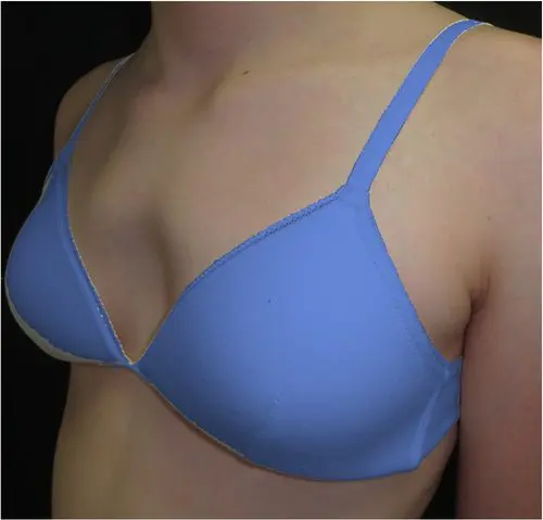
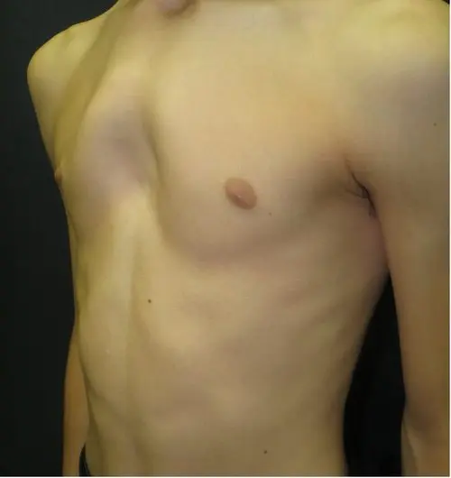
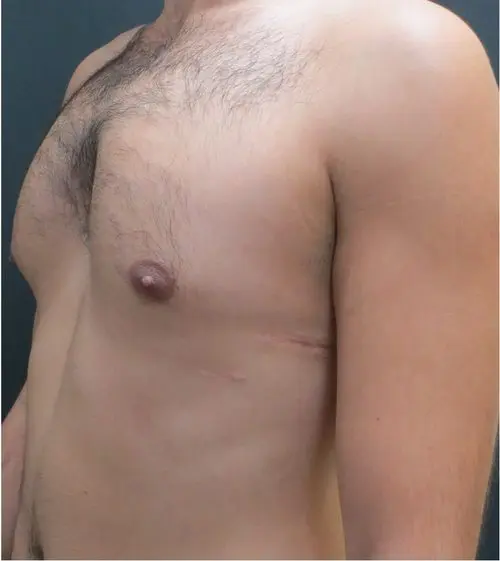
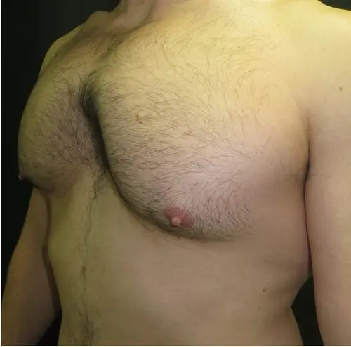
Pectus arcuatum shows relatively consistent shape across patients — the combination of upper protrusion and lower depression is the defining feature in all cases. The main variation is in severity, which often reflects the age and stage of skeletal development at presentation. Less pronounced deformity is more typical in younger pre-teen patients; more marked cases tend to present in adolescence or early adulthood.
How it is assessed
Pectus arcuatum is assessed clinically. Mr Hunt examines the full sternal contour, rib cage symmetry, and any associated features including connective tissue hypermobility and scoliosis. Because the deformity combines protrusion and depression, characterising the three-dimensional anatomy matters more than any single measurement.
Photographs in standard positions document baseline appearance. Where symptoms are present — chest discomfort, breathlessness, or reduced exercise tolerance — these are explored in detail and may prompt further investigation.
Surgery is the only option for structural correction of pectus arcuatum. Where correction is being considered, dedicated imaging and a formal surgical consultation are required before any recommendation is made.
When imaging is needed
CT chest with 3D reconstruction is requested in all patients being considered for surgical assessment. It characterises both sternal segments, costal cartilage morphology, and thoracic volume. Selective pulmonary function testing and cardiac review are arranged where there is functional concern.
Surgical assessment
Surgery is the only available option for correction of the deformity. A surgical consultation reviews CT anatomy, confirms suitability, and agrees the most appropriate approach for the individual patient.
Custom chest wall implant
For patients who wish to improve chest wall contour without skeletal correction, a custom-fitted implant may be considered as an aesthetic alternative to open surgery.
Implant surgerySurgical correction
Structural correction requires open or hybrid surgery tailored to the individual anatomy. Complex and revision cases are managed within a specialist pathway.
Treatment options
Treatment is tailored to the individual. Not every patient with pectus arcuatum requires intervention. The decision depends on the severity and nature of the deformity, functional symptoms, psychological impact, and the patient’s own priorities.
Observation & monitoring
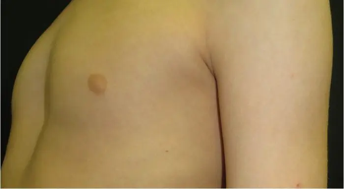
For patients with mild deformity and no significant functional symptoms, active monitoring is often the most appropriate initial approach. This involves periodic clinical review — typically yearly in adolescents — to track any change in the deformity through growth and to reassess the need for intervention over time.
Targeted exercise & postural retraining
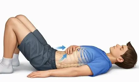
A structured exercise programme aimed at core strengthening, postural awareness, and chest wall muscle activation will not correct the underlying bony and cartilaginous deformity, but can improve functional capacity and reduce musculoskeletal discomfort. It is a useful component of both non-surgical management and surgical preparation and recovery.
External bracing
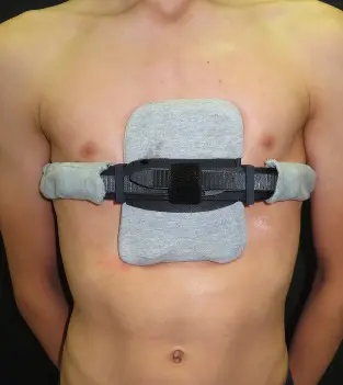
Expert assessment required. External bracing is typically not an option in pectus arcuatum. The combined protrusion and depression means that surface pressure alone risks displacing force onto the depressed sternal segment. It is considered only in the youngest and most skeletally flexible patients following specialist review.
Bracing has an established role in pectus carinatum by applying graduated pressure to the sternal protrusion. Its role in pectus arcuatum is more limited — the combined deformity means that addressing only the protrusion component risks displacing force onto the lower depressed segment. Where bracing is considered, it is custom-fitted and carefully monitored. It is most likely to be relevant in younger adolescents.
Surgery is considered when the deformity is symptomatic, the anatomy is unsuitable for non-surgical management, or when the psychological impact is significant and non-surgical options have been explored. Given the combined nature of the deformity, the operative approach requires individual planning.
Custom chest wall implant
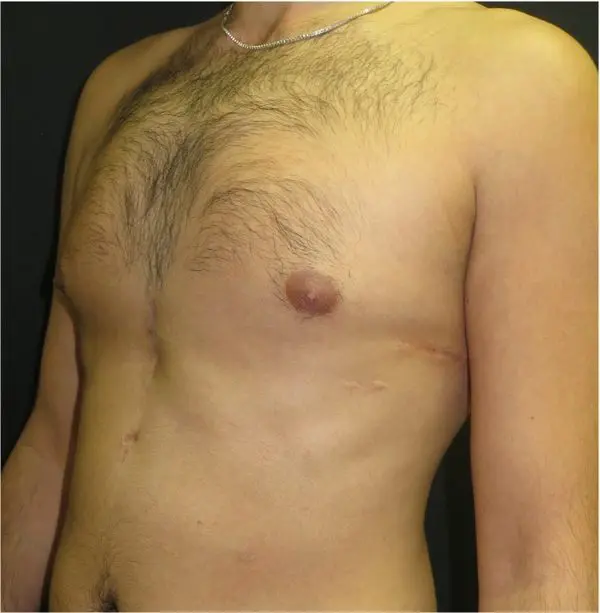
For patients who wish to improve chest wall contour without open skeletal correction, a custom-fitted silicone implant offers an alternative. Designed from CT data to match the individual anatomy, the implant fills the depressed lower sternal component to improve surface contour without a full Ravitch-type procedure or internal bar. Recovery is considerably shorter than for open correction.
Modified Ravitch procedure
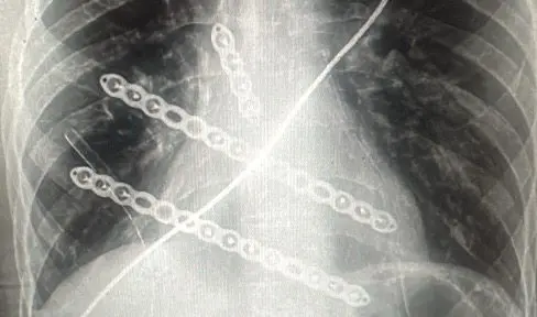
An open surgical technique in which the abnormal costal cartilages are resected and the sternum repositioned and stabilised. In pectus arcuatum, the procedure is modified to address both the upper protrusion and lower depression components in a single operation. Fixation is used to maintain the corrected sternal position while the cartilages remodel. It is a well-established approach for complex or combined deformities.
Hybrid correction
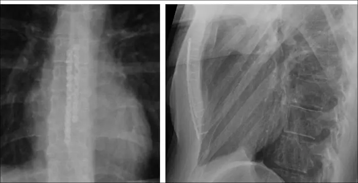
A combined approach using limited cartilage resection together with an internal bar to address the sternal contour. Hybrid techniques may allow correction of both the protrusion and depression components with less extensive dissection than a full Ravitch. Planning is based on detailed review of the CT anatomy. Not all patients are suitable and case selection is carefully considered.
Complex & revision surgery
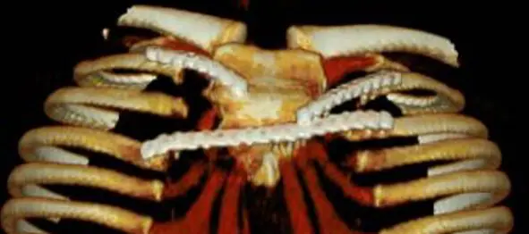
Some patients with pectus arcuatum present having had prior chest wall surgery elsewhere, or have a degree of complexity — co-existing rib flare, scoliosis, or connective tissue disorder — that requires a more individualised approach. The clinic has experience in complex and revision chest wall reconstruction, where standard approaches are modified to suit the individual anatomy.
Frequently asked questions
Is surgery always needed for pectus arcuatum?
No. Many patients with mild deformity and no significant functional symptoms are managed with observation and monitoring, particularly during adolescence. Surgery is discussed when the deformity is causing symptoms, is unlikely to improve with non-surgical measures, or is creating significant psychological distress. The decision is always made jointly with the patient after a full assessment.
Does pectus arcuatum get worse over time?
It often becomes more apparent during adolescent growth spurts. Once skeletal maturity is reached, the bony architecture is largely fixed, though symptoms may evolve with changes in physical activity, posture, or body composition. Regular clinical review during the growth years is helpful to track progression and inform the timing of any intervention.
How is pectus arcuatum different from pectus carinatum?
There are two fundamental differences. First, and most importantly, pectus arcuatum is a bony abnormality of the sternum itself — the sternal body is structurally abnormal in shape and orientation. Pectus carinatum, by contrast, is primarily a cartilaginous problem: the sternum is normal, but overgrowth of the costal cartilages pushes it forward. Second, pectus arcuatum produces both a protrusion of the upper sternum and an apparent depression of the lower segment, giving the characteristic wave or Z-shaped contour — whereas pectus carinatum involves forward protrusion only. These distinctions explain why the two conditions require different assessment approaches and why treatments effective for pectus carinatum, such as bracing, have a very limited role in pectus arcuatum.
Can bracing correct pectus arcuatum?
Not in the way bracing corrects pectus carinatum. Bracing works in pectus carinatum because the deformity is cartilaginous and relatively flexible — sustained surface pressure can gradually reshape the overgrown cartilages in growing patients. Pectus arcuatum is fundamentally different: the deformity is bony, involving the sternum itself, and the rigid bone does not respond to external pressure in the same way. Additionally, the combined protrusion and depression means that applying force to the upper sternum risks displacing load onto the depressed lower segment. Bracing has a very limited role and is considered only in exceptional circumstances in the youngest, most skeletally immature patients, and only following specialist assessment.
What is the recovery after surgery?
Recovery depends on the surgical approach used. Modified Ravitch procedures typically involve a hospital stay of two to four days, with a gradual return to normal activity over six to eight weeks. More complex procedures may require longer recovery. Detailed expectations, including restrictions on physical activity and follow-up arrangements, are discussed thoroughly in the pre-operative consultation.
Will my insurance cover treatment?
Coverage varies between insurers and depends on the clinical indication. Treatment for symptomatic or functionally significant pectus arcuatum is more likely to attract insurance support than purely cosmetic correction. The clinic’s administrative team can assist with pre-authorisation queries. Further details are available on the Fees & Funding page.
Arrange a specialist assessment
All patients are seen by Mr Ian Hunt personally. Consultations include a full clinical examination, review of any existing imaging and a clear, unhurried discussion of the options available.