Nuss Procedure
& Hybrid Surgery
The Nuss procedure is the established minimally invasive operation for pectus excavatum — using one or more shaped support bars to elevate the breastbone from within. For very asymmetric, complex or revision cases, a tailored hybrid approach draws on both minimally invasive and open techniques.
Surgery at the Pectus Clinic is consultant-delivered throughout. Every patient is assessed personally by Mr Ian Hunt, and the operative strategy is planned individually — no two chest walls are the same.
Surgical correction of pectus excavatum
Surgery is the established surgical treatment for pectus excavatum — appropriate when the deformity is significant, when it causes physical symptoms or marked distress, or when non-surgical options are unsuitable or have not achieved enough. The two operations offered at the Pectus Clinic for pectus excavatum are the Nuss procedure (minimally invasive repair, MIRPE) and hybrid chest wall surgery for more complex anatomy.
The decision to operate is never taken in isolation. It follows a structured assessment of the deformity itself, its effect on heart and lung function, the patient’s symptoms and goals, and — importantly — their age and stage of growth. For many younger patients a non-surgical programme is the right first step; for others, surgery is clearly the most appropriate option from the outset.
Mr Hunt performs the full range of chest wall operations, which means the recommendation made at consultation is based on what suits the individual chest — not on what a single-technique practice happens to offer. Where surgery is not the right answer, that advice is given just as directly.
At a glance
How this page works — the patient journey
The Nuss procedure elevates the sternum using one or more curved bars placed behind the breastbone through small keyhole incisions. Hybrid surgery adds selected open techniques for complex, asymmetric or revision deformity. Surgery is considered when the deformity is significant and symptoms, physiological impact or distress justify intervention.
Most patients return to normal daily activity and most non-contact exercise by around 12 weeks. The bars remain in place for approximately 3 years before planned removal under a separate short anaesthetic.
What is the Nuss procedure?
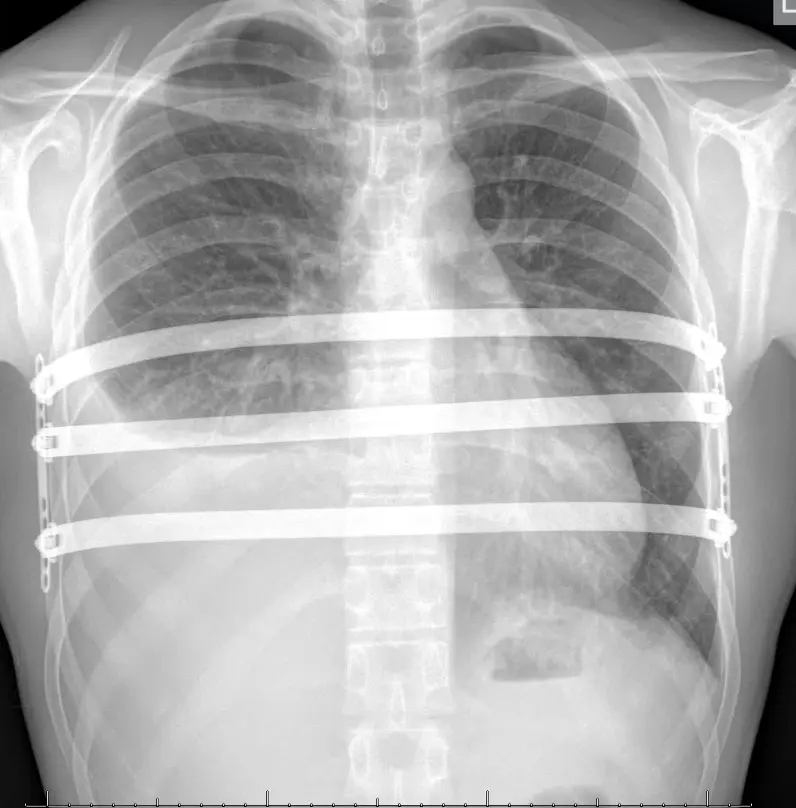
The Nuss procedure — minimally invasive repair of pectus excavatum (MIRPE) — corrects the deformity from within, without removing any cartilage or bone. It is performed through small incisions on either side of the chest under camera guidance.
In its basic form the operation follows four steps. In practice the technique has evolved considerably since it was first described — the refinements that make modern repair safer, more comfortable and more adaptable to individual anatomy are set out in The evolution of corrective pectus surgery below.
Assess & look across the chest
A small thoracoscopic camera is introduced so the inside of the chest can be inspected directly. The deepest point of the deformity, the path behind the sternum and the position of the heart are all assessed before anything is passed across.
Bar passed
A curved support bar, shaped to the individual chest, is passed across the chest behind the sternum under direct camera vision — the heart and pericardium kept in view throughout the retrosternal passage.
Bar rotated
The bar is inserted curve-down, then rotated. As it turns, its convex surface lifts the sternum and anterior chest wall forward into a corrected position in a single controlled movement.
Bar secured
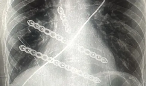
The bar is fixed to the chest wall with lateral stabilisers and sutures, and additional bars are added where planned. The bars hold the corrected shape while the chest wall remodels around them.
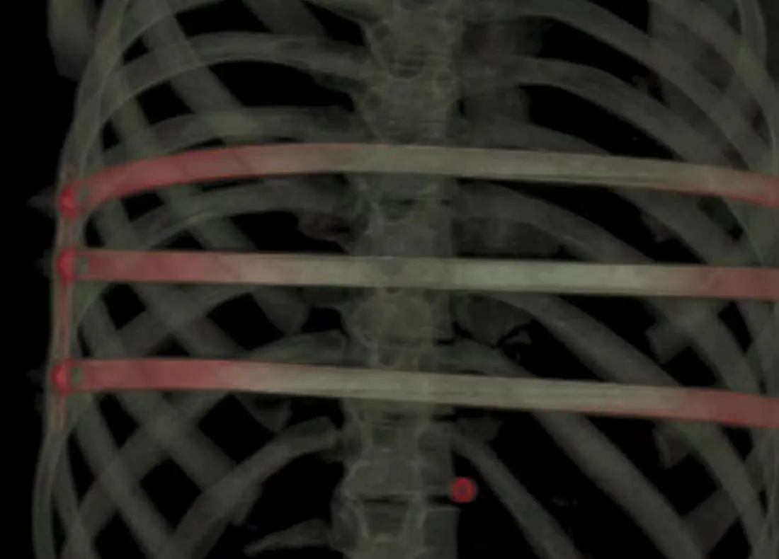
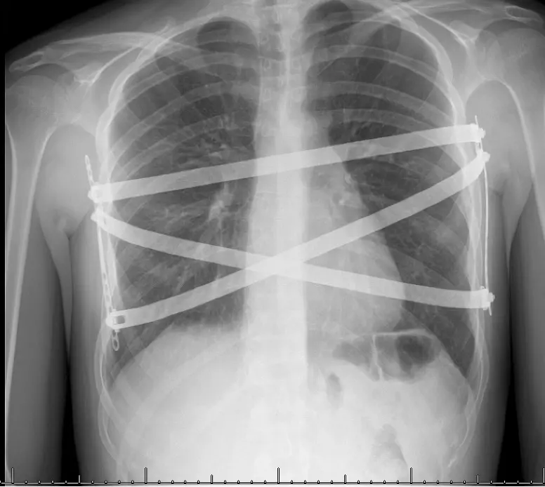
Depending on the shape, depth, length and symmetry of the deformity, one, two or three bars are used. Modern practice favours sharing the correction across several well-positioned bars rather than forcing it through a single point — this improves both the quality of correction and comfort afterwards.
Pain control is planned in advance and built into the operation itself. A combination of techniques is used — intercostal nerve cryoablation (freezing the chest-wall nerves under camera vision to switch off pain signals for weeks to months; temporary chest-wall numbness is expected and usually resolves, though a small area of altered sensation can occasionally persist), regional anaesthetic blocks such as paravertebral or erector spinae blocks, and patient-controlled analgesia (PCA) in the early post-operative period. Together these keep patients comfortable while substantially reducing the need for strong opioids.
Donald Nuss — origin of the operation
The procedure was developed by paediatric surgeon Donald Nuss in Norfolk, Virginia, who performed the first repairs in 1987 and published the landmark 10-year series in 1998. By avoiding cartilage resection altogether, it transformed pectus surgery and is now the most widely performed pectus operation worldwide.
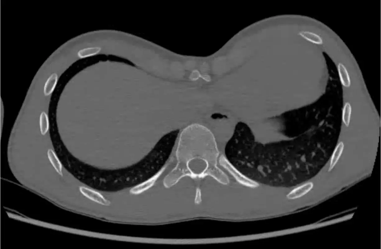
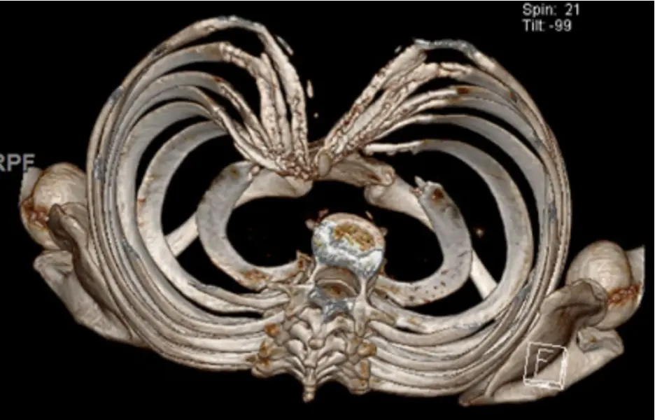
Matched CT slices at the same level of the chest, before and after a three-bar Nuss repair. The funnel deformity (Haller index 4.4) is elevated to a near-normal chest contour (Haller index 2.8), restoring space in front of the heart.
The evolution of corrective pectus surgery
The Nuss (or MIRPE) procedure performed today is based on the original technique first described in the 1990s, but now incorporates a great many refinements. Three decades of development — in imaging, instrumentation, fixation and anaesthesia — have steadily made it safer, more comfortable and capable of correcting deformities once considered suitable only for open surgery. The most important advances are summarised below.
Safer surgery, under direct vision
- Thoracoscopic guidance. The whole retrosternal passage is now performed under camera vision, with the heart and pericardium kept in view — an important factor in the operation's modern safety record.
- Sternal elevation. Lifting the sternum before the bars are passed — using a dedicated crane or hook device — is now considered mandatory, and is arguably just as important for safety: it opens the space behind the sternum and substantially reduces risk during dissection.
- Wider mediastinal dissection. A more deliberate, controlled dissection across the mediastinum allows the bar to be positioned exactly where it is needed rather than where it happens to pass.
- Pleural drainage & closure. Routine evacuation of air and, where needed, drainage has made post-operative recovery smoother and more predictable.
- Titanium bars. Modern bars are titanium rather than nickel-containing stainless steel — better tolerated, better MRI-conditional characteristics, and largely removing the problem of metal allergy.
Better, more comprehensive correction
- Multiple bars. Using two, three or more bars spreads the correcting force, allows a stronger and more distributed correcting force, and holds the shape far more reliably than a single bar ever could.
- Bar stabilisers & cross-fixation. Modern stabilisers — and securing bars to one another — have dramatically reduced bar displacement, the commonest early problem of the original technique.
- Lower bar placement. Because bars are now cross-secured to each other, they can be placed lower than the base of the sternum — reaching the part of the chest where much of the deformity actually lies, and allowing more comprehensive correction in suitable cases.
- Rib-flare control. In flexible chests, associated lower rib flare can be addressed at the same time using FiberTape sutures to draw the flared costal margins inwards.
- Post-operative CT assessment. Ideally, a chest CT post-operatively should be performed to demonstrate radiologically that the sternum and lower chest wall are well corrected, that no cardiac or lung compression remains, and to allow the post-surgery Haller Index to be calculated.
Comfort & complex cases
- Cryoanalgesia & regional blocks. Intercostal cryoablation and paravertebral / erector spinae blocks, alongside PCA, have transformed what was once a notoriously painful recovery.
- Hybrid techniques. Combining Nuss-style bar support with limited open work uses the advantages of both approaches — the chest-wall remodelling of the Nuss bars together with the direct sternal mobilisation of a mini-Ravitch.
- Extreme & very asymmetric deformity. These hybrid methods make it possible to correct markedly asymmetric chests, rigid adult chests and older patients who would previously have been turned away.
- Revision surgery. The same refinements underpin reliable correction of recurrent or failed previous repairs.
Hybrid chest wall surgery
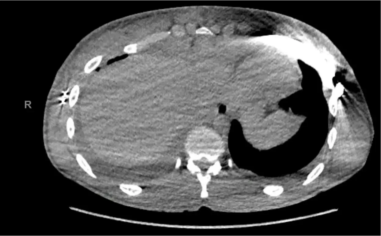
Not every chest is suited to a standard Nuss repair. Markedly asymmetric deformities, mixed excavatum–carinatum patterns, pectus arcuatum, very rigid adult chests and revision cases after previous surgery often need more than a bar alone can achieve.
Hybrid surgery combines elements of the minimally invasive and open approaches in a single tailored operation — for example, targeted reshaping or controlled division of abnormal costal cartilages, or a sternal osteotomy to correct rotation or angulation, combined with Nuss-type bar support to hold the corrected position while it heals.
The precise combination is planned from cross-sectional imaging before surgery and refined at operation. The aim is always the same: the best achievable correction with the least surgical trauma necessary to deliver it.
Mr Hunt has a specialist interest in complex and revision chest wall surgery, and accepts referrals of patients whose previous repair has recurred, displaced or produced an unsatisfactory result.
 Before
After
Before
After
When a hybrid approach is considered
Who is suitable for surgery?
Suitability depends on the severity and pattern of the deformity, its physiological and psychological impact, age and skeletal maturity, and the patient’s own goals. Surgery is a positive choice — it is never the default simply because a deformity exists.
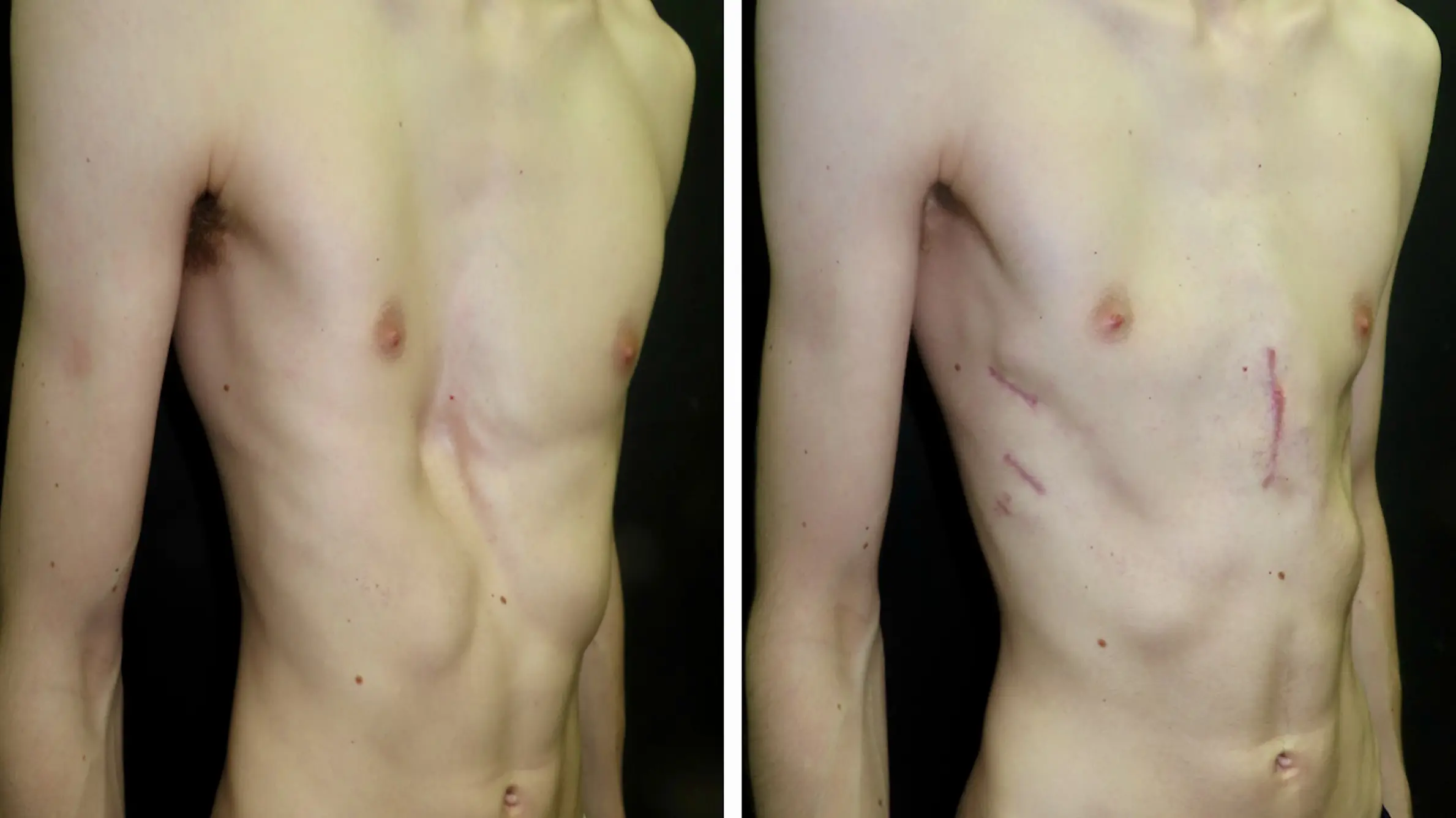
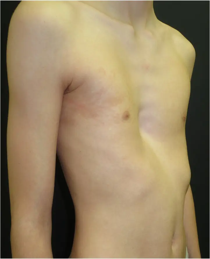
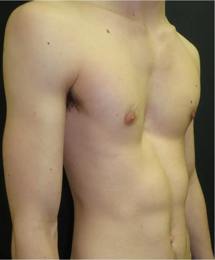
Most patients seen for surgery have a deformity of moderate to severe degree — often a visible funnel, with asymmetry or associated rib flare. The photographs alongside show a typical pectus excavatum before surgery and the same chest after minimally invasive correction.
The purpose of assessment is twofold: to confirm that surgery is genuinely the right route for that individual, and to plan the operation precisely around their anatomy, symptoms and goals.
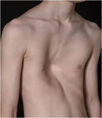
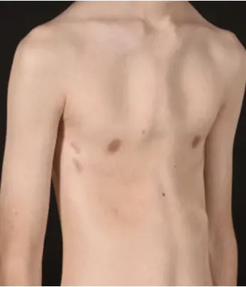
Pectus excavatum before, and after minimally invasive (Nuss) correction.
✓ Surgery is typically considered for
- Moderate to severe pectus excavatum — usually with a Haller index above ~3.25 or equivalent correction index
- Symptoms attributable to the deformity: exercise limitation, breathlessness, palpitations or chest discomfort
- Evidence of cardiac compression or displacement on imaging
- Significant impact on body image and quality of life
- Adolescents approaching or past the pubertal growth spurt — commonly from ~16 years
- Adults across a wide age range — including many previously told they were “too old” for repair
- Recurrence or unsatisfactory result after previous pectus surgery
✗ Surgery is less likely to be recommended when
- The deformity is mild and causing neither symptoms nor significant distress
- A younger, flexible chest is likely to respond well to vacuum bell therapy first
- Growth is at a stage where operating now would risk recurrence — timing matters
- Expectations are primarily cosmetic-perfectionist rather than realistic correction
- Significant medical comorbidity makes general anaesthesia inadvisable
Surgery is considered for patients with significant pectus excavatum — from adolescents approaching the end of their growth to adults across a wide age range. The key factors are the severity of the deformity, the presence of symptoms, and the patient's goals. No recommendation is made without a full consultation and imaging review.
Assessment and surgical planning
Every surgical patient follows a structured assessment pathway. Its purpose is twofold: to confirm that surgery is the right treatment for the individual, and to plan precisely how the correction will be performed. Assessment is consultant-led throughout — not reduced to a single measurement.
Medical photography. Standardised clinical photographs are taken at the outset to record the starting appearance and allow objective comparison after correction.
Imaging. A low-dose CT scan of the chest with 3D reconstruction defines the depth, symmetry and extent of the deformity, the degree of sternal rotation, and the relationship of the breastbone to the heart. From this the Haller index and correction index are calculated, and the number, length and shape of the bars are planned.
Echocardiogram. An echocardiogram (heart ultrasound) assesses whether the depressed sternum is compressing or displacing the heart, and screens for the valve and filling effects that can accompany more severe pectus excavatum.
Cardiac MRI. Where a clearer picture of the cardiac effect is needed, a cardiac MRI provides a dynamic, radiation-free view of how the sternum indents and displaces the heart through the cardiac cycle — particularly useful in symptomatic patients and in building the evidence base for insurance funding. The short loop alongside is a cardiac MRI in a patient with pectus excavatum.
Lung function and cardiopulmonary exercise testing (CPET). Lung function tests document any effect of the deformity on breathing. Where exercise capacity is in question, cardiopulmonary exercise testing (CPET) measures how the heart and lungs perform together under exertion — often the most useful objective measure of functional impact, and valuable both for surgical decision-making and for pre-operative documentation. These cardiac and respiratory investigations are arranged within Mr Hunt’s specialist network.
Anaesthetic review. Finally, every patient is assessed before admission by a consultant anaesthetist experienced in chest wall surgery, with the pain-management plan agreed in advance.
The planning checklist
Assessment is planned individually. Most patients will need a formal CT scan for Haller Index measurement, cardiac imaging (CMR or echocardiogram) to document compression, and in some cases cardiopulmonary exercise testing (CPET). All investigations are arranged and reviewed before any surgical decision is made.
NHS funding criteria for pectus surgery
Since 2023, NHS England funds surgery for pectus excavatum only where there are very severe, objectively documented physiological symptoms. A referred patient is generally considered when there is:
- a Haller Index >3.25 on CT scanning (a formal measure of the depth of the depression, documented on CT imaging), together with
- objective evidence of a functional effect — for example compression or displacement of the heart on cardiac MRI, a cardiac arrhythmia or other cardiac symptoms, significant breathlessness or exercise limitation, or swallowing difficulty (dysphagia).
Every referral is reviewed independently by a National Pectus MDT (a multidisciplinary team meeting — a panel of different specialists who consider each case together), which decides whether a patient qualifies. Those accepted are treated at one of the national pectus centres. Pectus carinatum and purely cosmetic concerns are not funded.
Mr Hunt can advise whether you are likely to meet these criteria and help arrange the appropriate referral.
The operation — what actually happens
A primary Nuss procedure is carried out under general anaesthetic as a keyhole (minimally invasive) operation, typically taking around four hours. A hybrid operation takes longer, adding a limited incision at the front of the chest. The stages below describe a standard Nuss repair; the surgical images are shown blurred and can be revealed with a click.
Anaesthetic & preparation
You are admitted on the day of surgery and anaesthetised. The chest is positioned and marked, identifying the deepest point of the deformity and the planned path of the bars. Antibiotics are given. A regional anaesthetic block — typically a paravertebral block, where local anaesthetic is placed alongside the nerves as they leave the spine to numb the chest wall — is usually used as part of the pain-control plan.
Small lateral chest incisions
Small incisions are made on each side of the chest. A thoracoscopic camera is introduced through one of them, so the inside of the chest — and the position of the heart — is seen directly throughout. This is video-assisted thoracoscopic surgery (VATS).
Cryotherapy — freezing the nerves
Where used, the intercostal nerves running along the ribs are frozen under camera vision. This temporarily switches off pain signals from the chest wall for weeks to months, substantially reducing the need for strong painkillers and helping patients breathe, move and go home sooner.
Sternal elevation
Typically a very small incision is made at the front of the chest, closest to the deepest part of the deformity. Using a specialist lifting instrument, the sternum (breastbone) is raised away from the heart, creating room to work safely beneath it.
In a hybrid repair this midline incision is lengthened. The costal cartilages holding the breastbone down are divided to release the sternum, and where necessary the sternum itself is partially cut, also releasing it — allowing a stiff or markedly rotated chest to be mobilised before the bars are placed.
Mediastinal dissection & support sutures
Under direct camera vision, a careful dissection is made across the mediastinum — the space behind the sternum and immediately in front of the heart — to create a controlled path for the bars. Support (‘hammock’) sutures are then placed around the ribs so that each bar comes to sit on them, rather than stripping the intercostal muscle. This supports the bars and helps reduce the risk of later displacement.
Bars inserted & rotated
Two or more bars — pre-shaped at the bedside, meaning each is bent to match the individual chest contour during the operation — are passed across behind the sternum and then rotated to lift the chest into a corrected position. The correction is immediate.
Bars secured & drains placed
The bars are fixed to the chest wall with lateral stabilisers and sutures. One or two pleural drains are placed, and the lung is seen to re-expand with the help of the anaesthetist.
Wound closure & recovery
The wounds are closed with absorbable (internal) sutures and dressed. In the recovery area a chest X-ray confirms the lung has fully re-expanded, and a patient-controlled analgesia (PCA) pump is started for comfort in the first day or two.
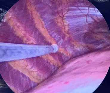
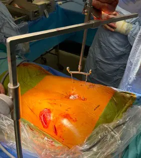
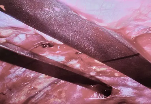
Surgical videos from our specialist network
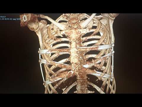
Mr Hunt works alongside Mr Joel Dunning, an excellent Consultant Thoracic Surgeon and valued colleague within his specialist network. The featured video shows Mr Dunning’s modern approach to a particularly challenging pectus excavatum, and his YouTube channel offers a library of detailed, narrated operative videos — a valuable resource for patients who want to understand exactly what the operation involves.
Further operative videos:
- The Nuss procedure using titanium (Park Prime) bars and bridges
- How a CT scan is measured for pectus excavatum
- The mini-Ravitch operation
- A redo (revision) Nuss procedure
These are external videos hosted on YouTube and contain surgical footage.
Pain control — a modern, planned approach
The Nuss procedure has a reputation for being painful — historically with good reason. Modern multimodal pain management has changed this substantially, and a clear pain plan is agreed with every patient before surgery.
Regional paravertebral block. A paravertebral block is usually placed at the time of surgery: local anaesthetic is injected alongside the spinal nerves as they emerge, just outside the chest cavity, numbing the chest wall on the operated side. It provides strong, targeted pain relief in the first hours and days, reducing the amount of opioid needed and helping patients breathe deeply and move early. Additional local anaesthetic is also infiltrated directly around the incisions during the operation for added comfort as the block wears off.
Cryotherapy (cryoanalgesia). Where appropriate, the intercostal nerves are frozen under camera vision during surgery. This temporarily switches off pain signalling from the chest wall for weeks to months while the nerves regenerate — substantially reducing painkiller requirements and, in published series, shortening hospital stay.
Multimodal medication. These regional techniques are combined with a patient-controlled analgesia (PCA) pump in the first day or two and regular oral painkillers, alongside anti-sickness (antiemetic) medication as required. Layering different drug types keeps patients comfortable while avoiding reliance on any single class and limiting heavy sedation.
Structured step-down. Pain medication is then reduced in a planned, staged way after discharge, with clear written guidance and direct access back to the team if comfort is not as expected.
What patients can expect
Recovery after Nuss and hybrid surgery
Recovery is staged and predictable for most patients. On average it takes about four to six weeks to recover from the surgery, and up to twelve weeks before returning to full physical activity and sport. The framework below is a typical course — individual guidance is given at each follow-up review, and hybrid or revision operations may run slightly more slowly.
Days 0–5 — typically around 3 days
Early mobilisation is the single most important part of early recovery — walking starts the day after surgery, helping reduce the risk of blood clots and chest infection.
At home — the first 12 weeks
Weeks 1–4
- Sleep on your back; avoid sleeping on your side.
- Avoid sudden or excessive bending and twisting of the upper body.
- Keep good posture — bend at the hip rather than slouching; no backpacks for the first 3 months.
- Avoid lifting or carrying heavy weights that make you strain — this protects against bar movement.
- Walking and deep-breathing exercises (incentive spirometry) are encouraged.
- Regular painkillers are usually needed; most return to desk-based work or studies around 4–6 weeks.
- Wound dressings are removed about 7–10 days; you can shower but keep the wounds dry.
- Feeling off your food, tired or constipated is common — eat well and keep moving gently.
Weeks 4–8
- Increase your activity, including aerobic exercise such as jogging, cycling and swimming.
- You can lift and carry more, but continue to avoid heavy weight-lifting and straining.
- Painkillers are typically taken as required rather than regularly.
- The first follow-up review is around 4 weeks; after this most patients are able to travel, including by air.
Weeks 8–12
- Build steadily back towards normal daily activity and non-contact exercise.
- By around 12 weeks most patients have returned to normal daily activity and most non-contact exercise. Contact, collision and heavy-impact activities (rugby, martial arts, boxing) are discussed individually and usually deferred a little longer.
- A 12-week review confirms progress. The chest may stay a little numb if cryotherapy was used.
Contact the ward or the Pectus Clinic — or attend A&E if urgent — if you develop: a fever; a persistent cough; worsening chest pain, especially on deep breaths; sudden breathlessness; redness, swelling or discharge from a wound; any chest injury causing severe pain; or a sudden change in the shape of your chest with pain (which can signal bar movement).
Most patients are discharged 3–5 days after surgery and return to normal daily life and non-contact activity within around 12 weeks. Contact and collision sports require individual discussion. The bars remain in place for approximately 3 years before planned removal.
Risks and complications
The Nuss procedure is a major operation, but in fit patients and in specialist hands the risk of a serious or life-threatening complication is very low. It is important to be aware that, although only rarely reported, injury to the heart has caused death worldwide. The main complications are grouped below by how often they occur; all are discussed openly at the consent consultation, and tailored further for complex or revision cases.
Common · usually minor
- Chest pain, controlled with painkillers and settling over a few weeks
- Numbness of the chest or around the wounds — usual, and often for some months where cryotherapy is used; a small area can be permanent
- Temporary constipation, reduced appetite, tiredness and some weight loss
- Bruising and swelling around the wounds
- In slim patients, the bar can sometimes be felt
Uncommon
- Bleeding (around 1–2%) — occasionally a collection of blood (haemothorax) needing a drain, or rarely a return to theatre
- Pneumothorax — air around the lung if it does not fully re-expand; fewer than 1% need a drain
- Pleural effusion — a collection of fluid around the lung; usually monitored on X-ray and only occasionally drained
- Atelectasis — small areas of lung not fully expanding (micro-collapse), which can cause a feeling of breathlessness and improves with breathing exercises and physiotherapy
- Bar displacement — reduced by modern fixation techniques, stabilisers and cross-securing of bars; published rates vary and the risk is greatest in the first six weeks; repositioning is occasionally needed
- Wound infection or seroma — usually settle with dressings and antibiotics
- Differences in scar healing (wide, thickened or, rarely, keloid)
- Chronic, usually mild bar-related discomfort
Rare but serious
- Pericarditis or fluid around the heart — usually settles with anti-inflammatory treatment; very rarely drained
- ‘Buffalo-chest’ pneumothorax — because the bar passes across the chest, the two lung cavities can remain connected (a ‘buffalo chest’), so a collapsed lung can rarely become a both-sided collapse, causing sudden chest pain and/or collapse and needing emergency decompression
- Infection of the bar itself — may require the bar to be removed
- Injury to the heart or major vessels during bar passage — the most serious risk; made extremely rare by camera guidance and sternal elevation, but rare deaths have been reported worldwide
- Arm pain, numbness or weakness (Thoracic outlet syndrome). Thoracic outlet syndrome is a rare complication after Nuss surgery caused by compression of the nerves or blood vessels at the thoracic outlet, resulting in arm pain, numbness, weakness, or swelling.
- Recurrence of the deformity after the bar is removed (under 10%) — and usually a slight dip rather than a true recurrence
Results of Nuss and hybrid surgery
Published international experience of the Nuss procedure now spans more than 25 years and many thousands of patients. In experienced specialist centres, outcomes are consistently favourable.
Beyond the numbers, the change patients describe most often is simpler: standing differently, swimming without a t-shirt, no longer thinking about their chest every day. For symptomatic patients, published studies and registry data report improvements in breathlessness and exercise capacity following correction of significant deformities, though the strength of evidence varies; the RESTORE trial is expected to provide more robust data.
Results from hybrid and revision surgery are individually assessed — by definition these are more complex chests — but careful planning means expectations can be set realistically before any decision is made.
For most patients with significant pectus excavatum, surgery produces a durable improvement in chest contour and, in symptomatic patients, reported improvements in breathlessness and exercise capacity. Individual results depend on the severity and type of deformity, surgical approach, and recovery. The RESTORE trial is expected to provide the most robust published evidence to date.
The RESTORE Trial
Since NHS England stopped routinely funding pectus surgery, a major national study has set out to settle the question with high-quality evidence. The RESTORE Trial is an NIHR-funded, multicentre randomised controlled trial — the first of its kind — comparing corrective surgery with conservative (non-surgical) management for severe pectus excavatum. Led from South Tees Hospitals, it recruited 200 patients across around twelve UK centres, measuring the effect of surgery on cardiopulmonary function and quality of life. Randomised recruitment is complete, follow-up is ongoing, and an observational cohort remains open; the results are expected to inform future NHS funding decisions.
Mr Hunt supports the trial’s aims. His colleague Mr Joel Dunning, Consultant Cardiothoracic Surgeon at South Tees and the trial’s lead surgeon and chief investigator, introduces the study in the short video below.
Watch Mr Dunning explain the RESTORE Trial → About the trial (South Tees NHS) →
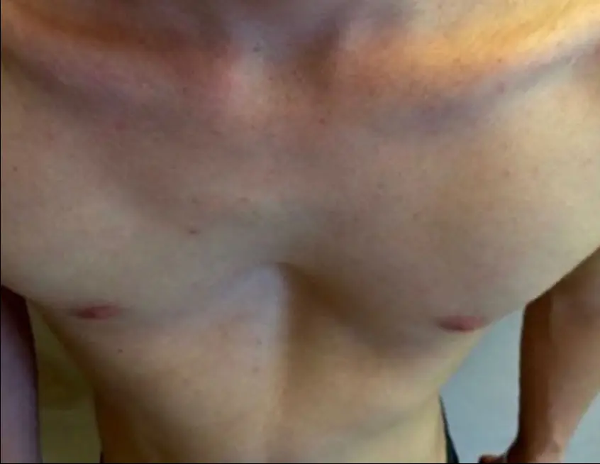
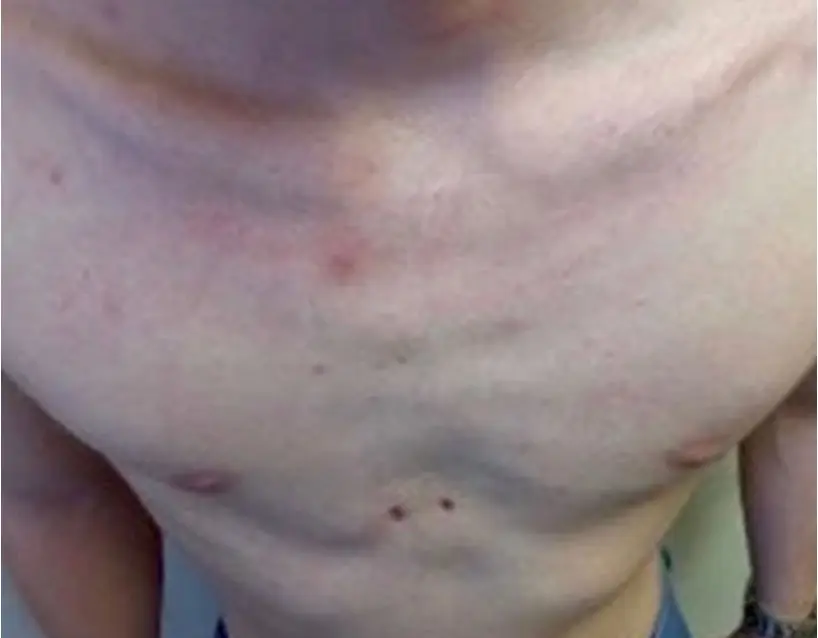
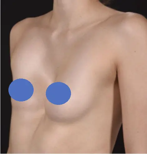
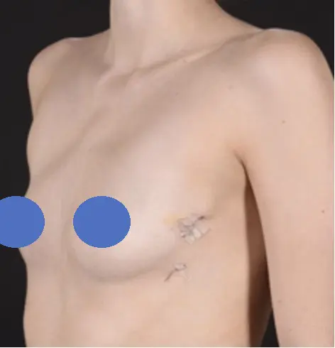
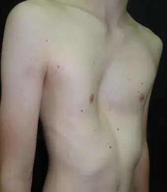
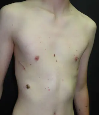
Before & after
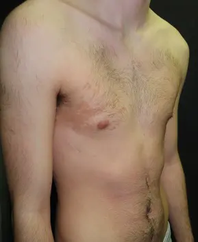
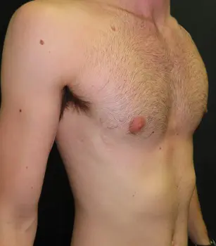
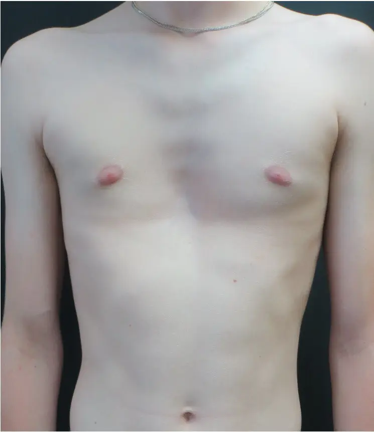
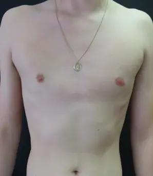
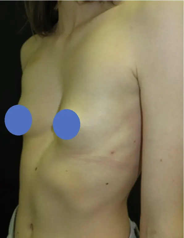
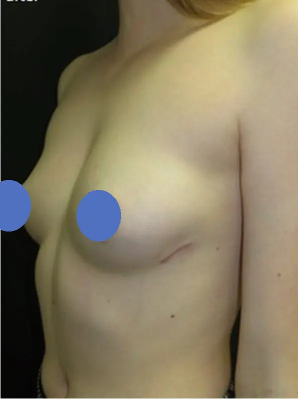
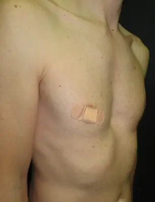
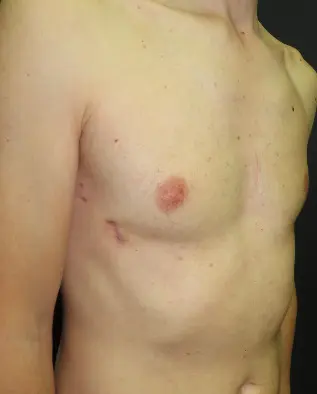
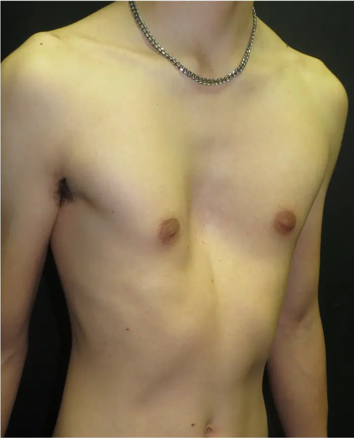
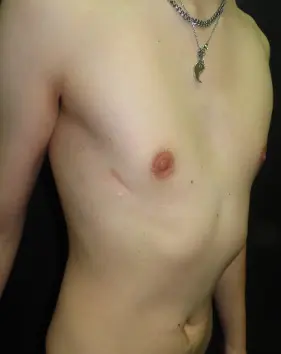
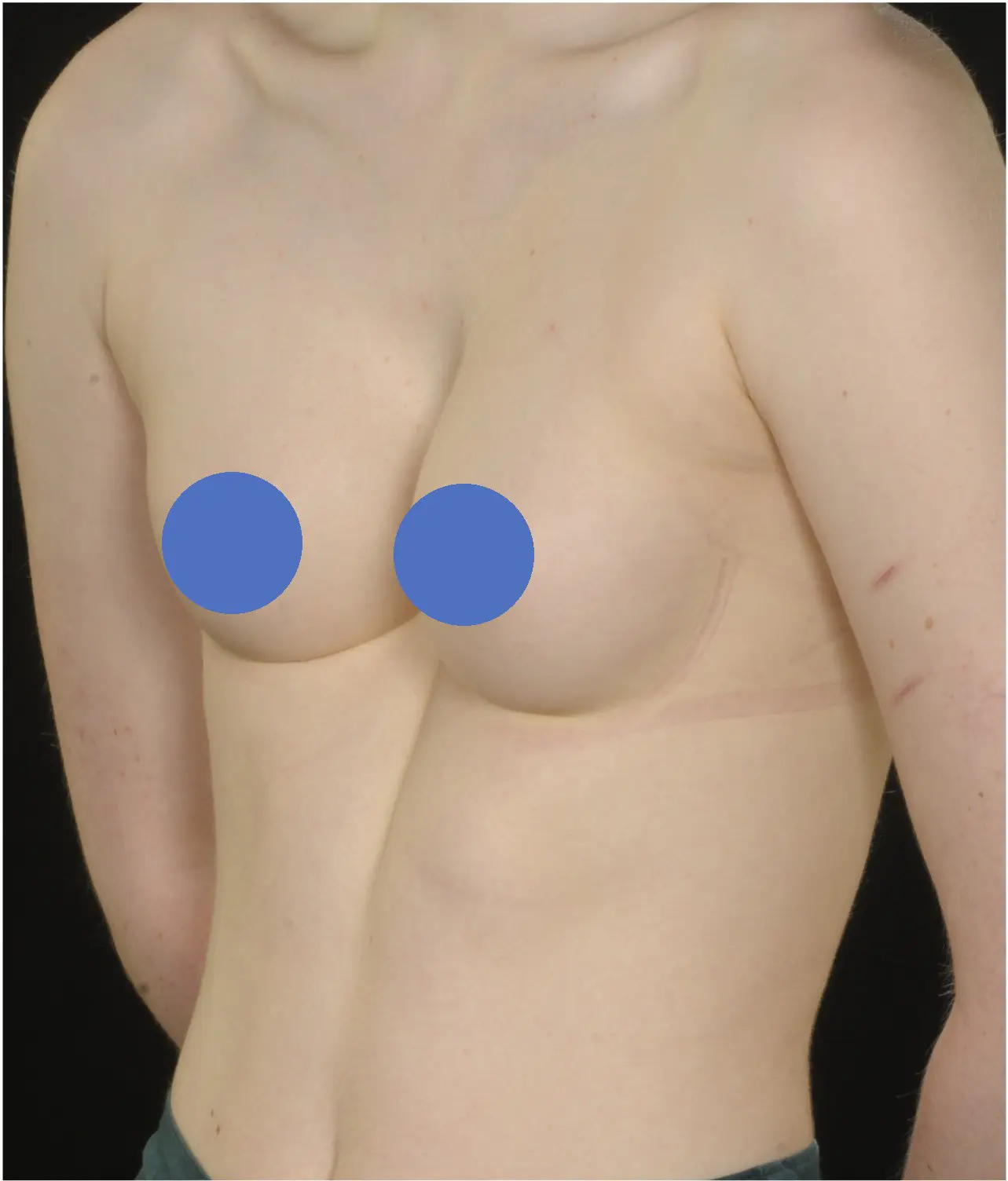
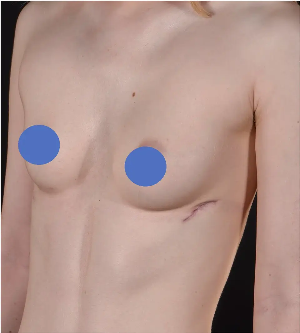
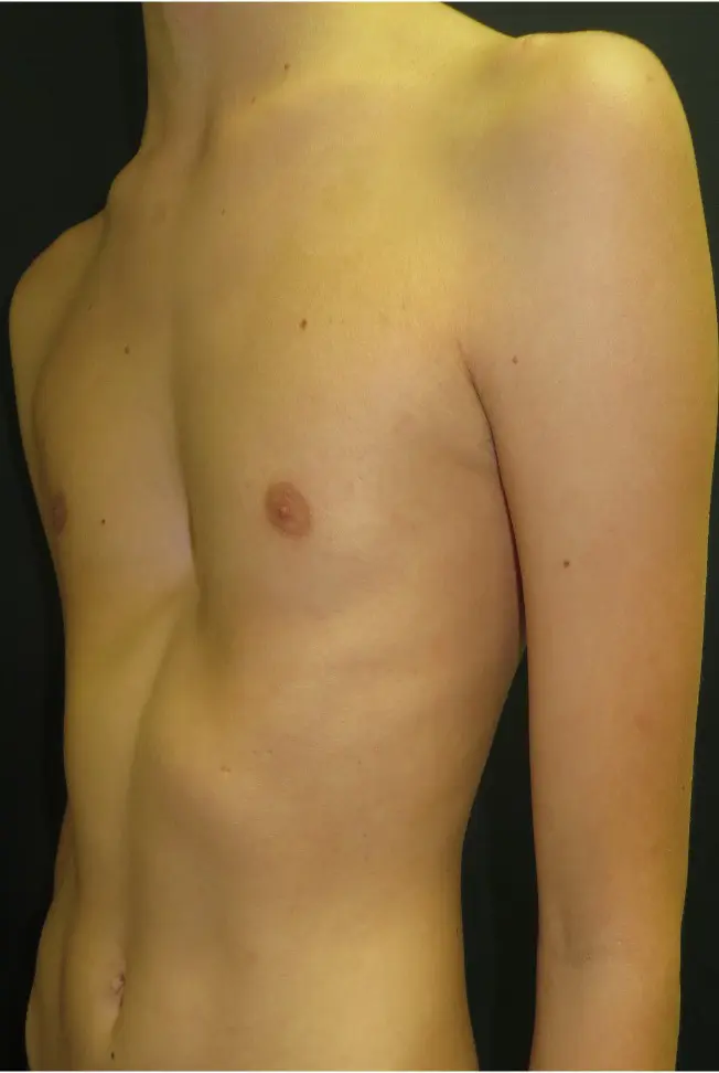
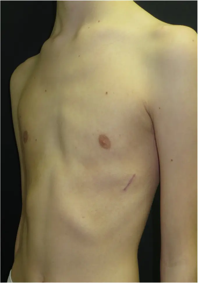
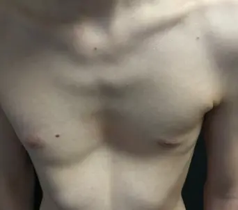
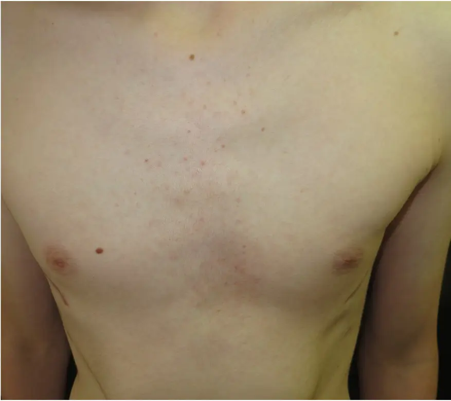
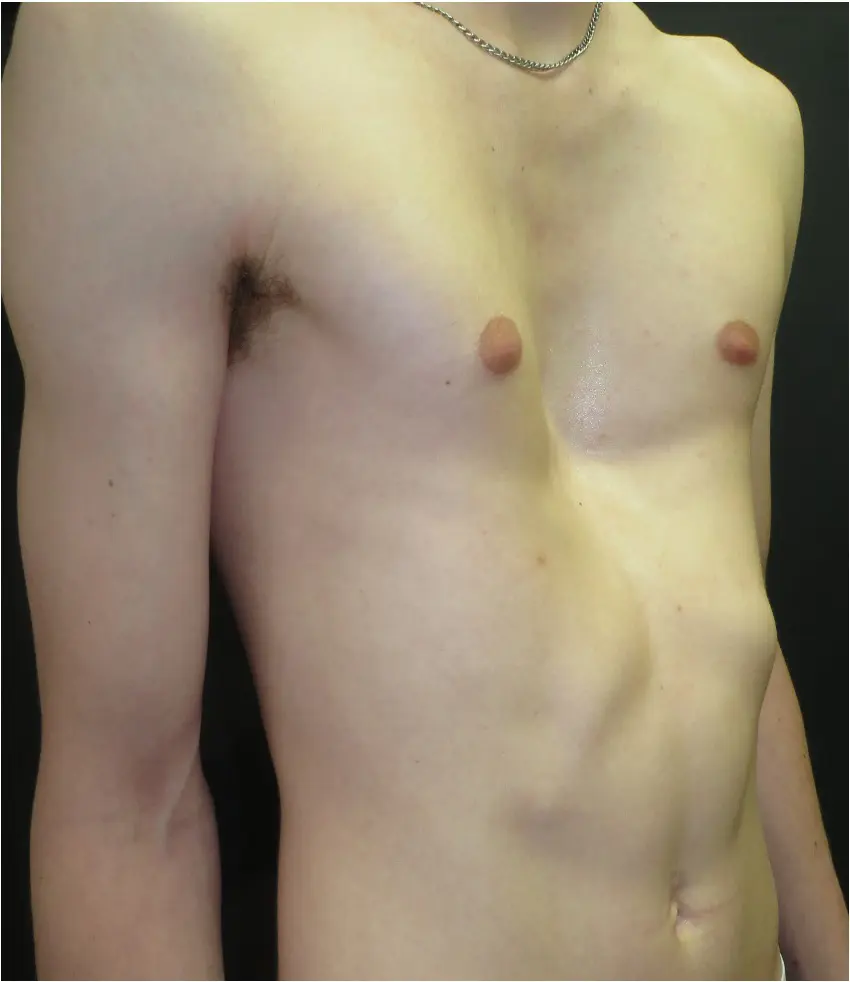
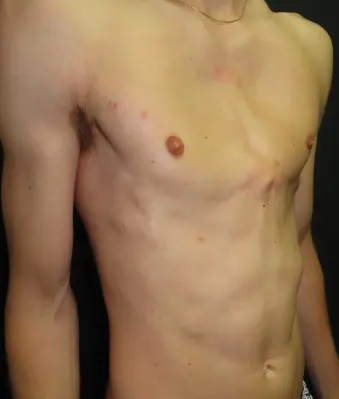
Clinical photographs from the Pectus Clinic surgical practice, shared with patient consent for educational and clinical illustration. Results reflect individual cases — outcomes vary and are discussed in detail at consultation.
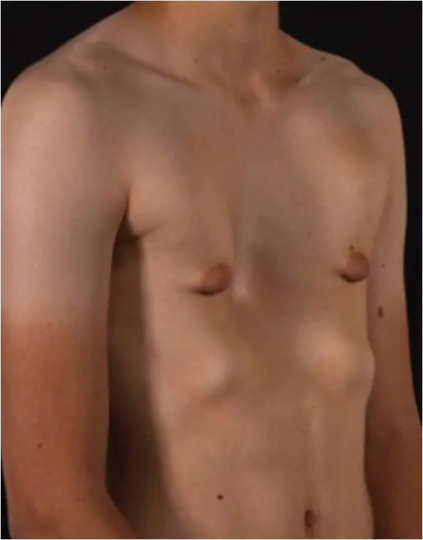 Before
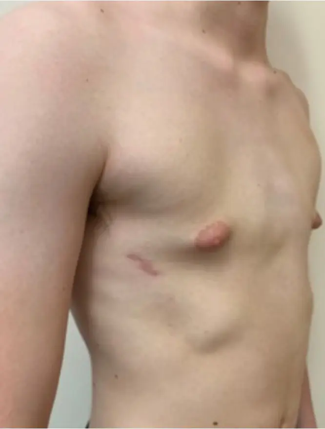 After
After
 Before
 After
After
 Before
 After
After
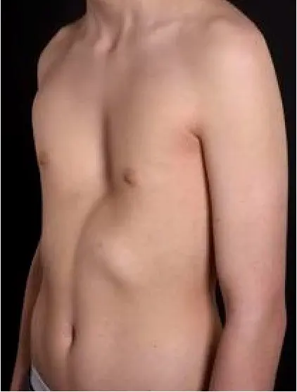 Before
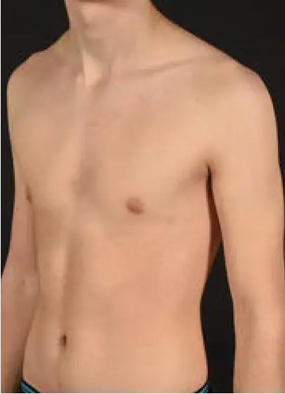 After
After
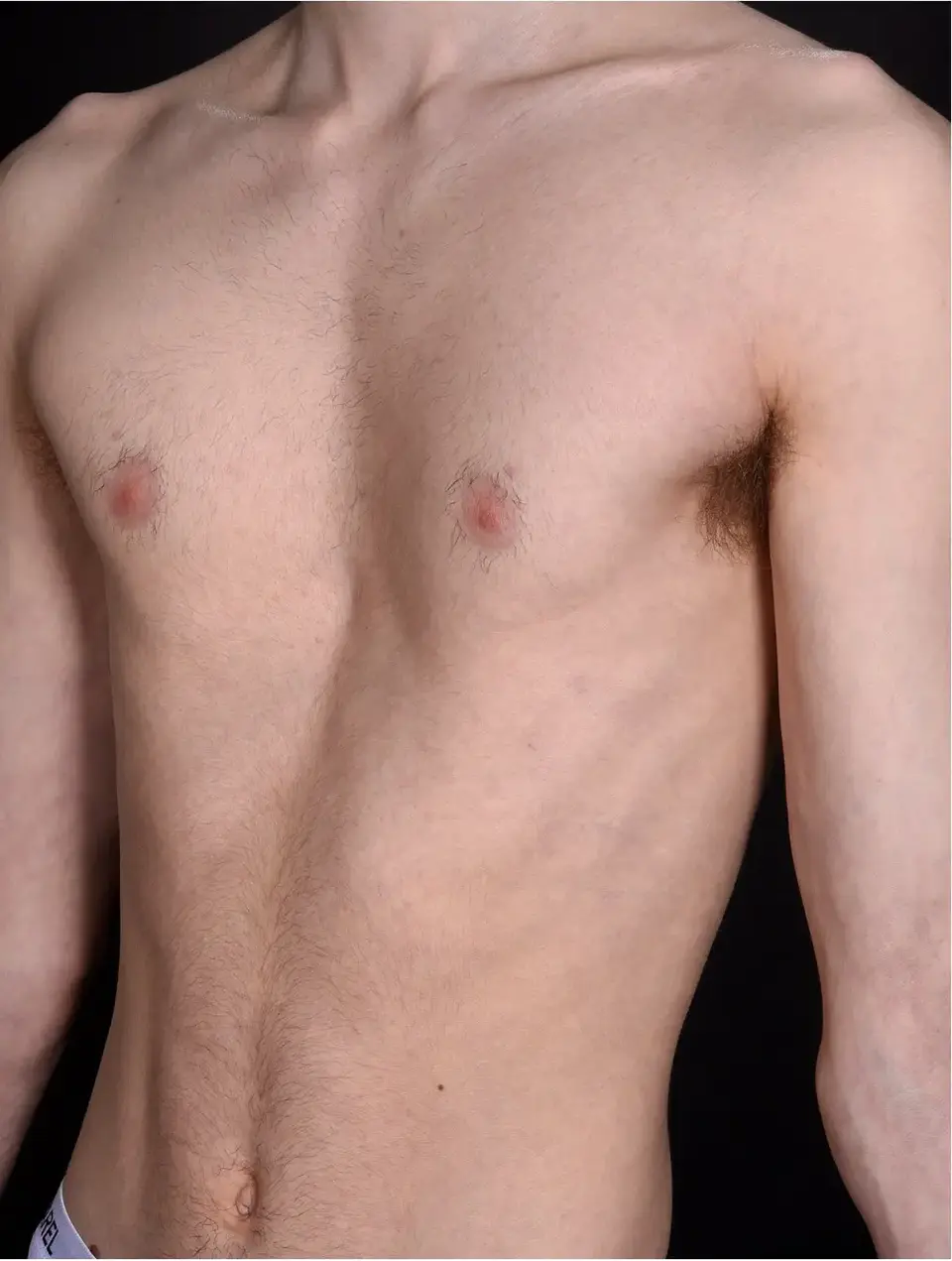 Before
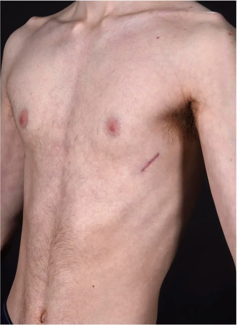 After
After
 Before
 After
After
 Before
 After
After
 Before
 After
After
 Before
 After
After
 Before
 After
After
 Before
 After
After
 Before
 After
After
 Before
 After
After
 Before
 After
After
 Before
 After
After
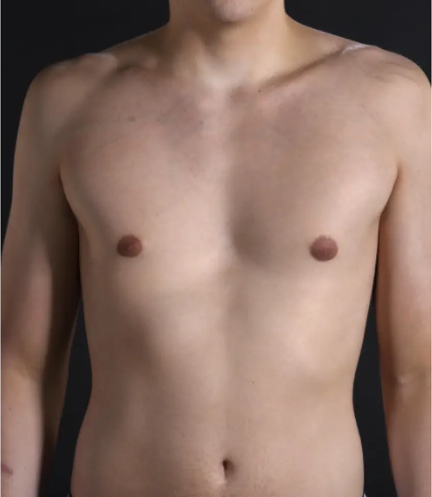 Before
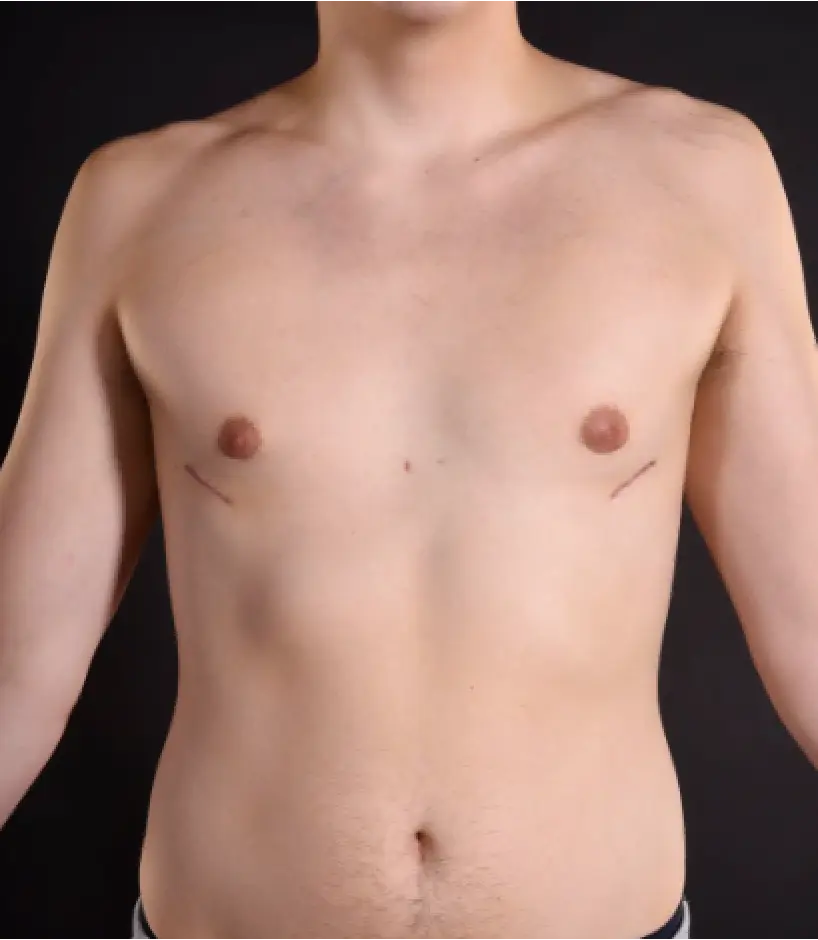 After
After
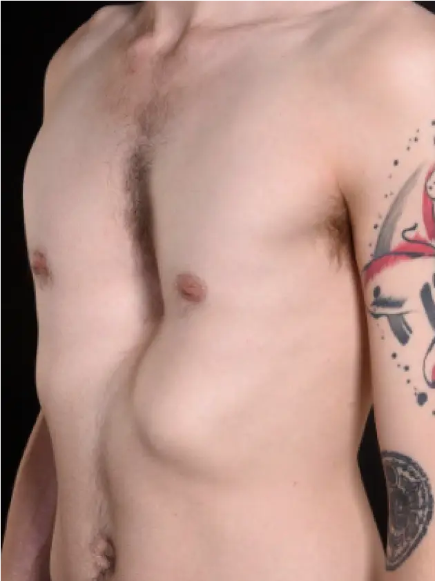 Before
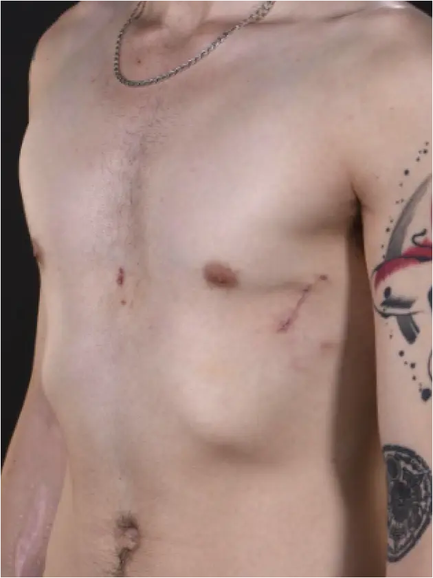 After
After
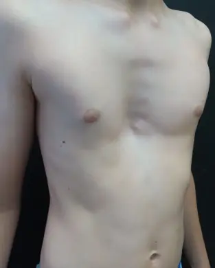 Before
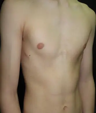 After
After
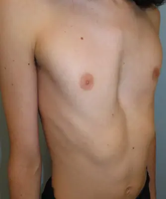 Before
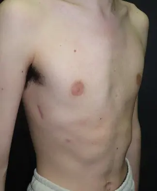 After
After
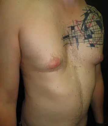 Before
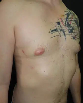 After
After
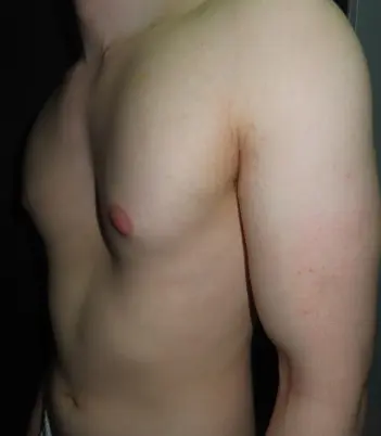 Before
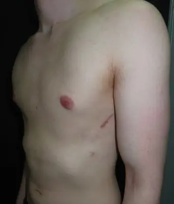 After
After
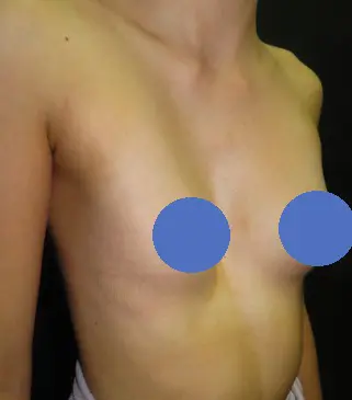 Before
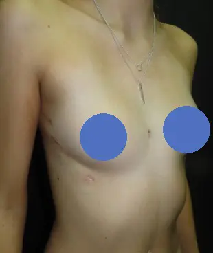 After
After
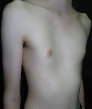 Before
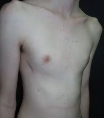 After
After
 Before
 After
After
Bar removal
The support bars are temporary. Once the chest wall has remodelled into its corrected shape — usually after around three to four years — the bars are removed at a planned second operation.
Bar removal is a much smaller procedure than the original repair: it is performed under general anaesthetic through the original lateral incisions, typically as a day case or with a single overnight stay, and recovery is measured in days rather than weeks.
The corrected chest shape is maintained after removal in the great majority of patients. Mr Hunt also performs bar removal for patients whose original surgery was carried out elsewhere — including overseas — and removal of bars that have become symptomatic or displaced.
Costs and funding for pectus surgery
Pectus surgery is funded in three main ways in the UK: NHS treatment (through the national NHS pectus programme, which applies strict assessment criteria before a patient is accepted for surgery), private medical insurance, and self-pay. The position is explained clearly at consultation so there are no surprises.
Yes — but only for severe cases meeting defined criteria, and through a nationally commissioned pathway. NHS England funds pectus excavatum surgery for patients with very severe, objectively documented physiological impairment. The pathway is: GP or specialist referral → investigation to demonstrate genuine cardiorespiratory effect → independent case review at a national pectus MDT → if accepted, surgery at a designated national centre. Pectus Matters provides up-to-date guidance on accessing NHS pectus surgery across all four nations.
NHS criteria for pectus surgery (England) (criteria may be updated — check current commissioning policy):
Access across the four nations (source: Pectus Matters, 2025)
The specific criteria and evidence requirements are discussed honestly at consultation. Where an NHS route is realistic for a patient, the team will explain what investigations are needed and how to support a referral. Many patients with significant functional impairment are not aware that NHS surgery is a possibility.
Private medical insurance. Many insurers fund pectus surgery where there is documented functional impact — symptoms plus objective findings on imaging, echocardiography or lung function testing. The assessment pathway at the Pectus Clinic is designed to provide exactly this evidence, and the team is experienced in supporting pre-authorisation applications. However, PMI cover for pectus surgery varies significantly between insurers — and within the same insurer, between individual policies. There is no guarantee of cover, and some policies exclude congenital or musculoskeletal conditions, impose benefit limits that do not reflect the true cost of complex chest wall surgery, or require specific pre-authorisation criteria that differ from clinical best practice. It is strongly advisable to seek written pre-authorisation before committing to surgery, and to check both surgical and anaesthetic cover, as these are sometimes assessed separately.
Self-pay. For self-funding patients, a clear written quotation is provided after consultation, covering the surgical, anaesthetic and hospital fees and follow-up; bar removal in due course is quoted separately. As a guide, a surgical package is approximately £20,000. The package includes all hospital, surgical and anaesthetic fees, and covers routine follow-up and indicated chest X-rays within the first year, as set out in the written quotation.
Nuss & hybrid surgery — FAQs
The first days after surgery are the most uncomfortable, and this is managed with a planned combination of techniques — including, where appropriate, intercostal cryoablation performed during the operation itself. Most patients are comfortable on oral medication by discharge and off regular strong painkillers within two to three weeks. Pain is taken seriously at every stage: it should always be controlled and improving.
Age alone is rarely a barrier. The procedure is well established in adults, including those in their 30s, 40s and beyond, though the technical approach is typically more demanding than in adolescents. Adult chests are stiffer, which usually means more bars, a more robust sternal elevation, and sometimes a hybrid component to release the costal cartilages. Recovery may take a little longer and pain management is generally more intensive. The decision rests on the severity of the deformity, the patient’s symptoms and goals, overall fitness, and the findings on imaging — not a fixed age threshold.
A standard Nuss repair leaves a small scar on each side of the chest, positioned laterally where they are inconspicuous — there is no scar on the front of the chest. Hybrid operations may involve an additional anterior incision, which is discussed and planned with you beforehand.
Most patients are aware of the bars at first, particularly with certain movements, but this fades and the bars are not visible from outside in the vast majority. Slim patients may feel the bar ends at the sides of the chest. Day to day, most patients live normally with the bars in place.
The bars may trigger airport metal detectors — patients are given a letter confirming the implant for travel. Modern pectus bars are MRI-conditional: scans can generally be performed safely, though the radiology team should always be told about the implant beforehand so the correct protocol is used.
Walking starts the day after surgery and builds steadily. Non-contact exercise — running, swimming, cycling, gym work — resumes in a graded way from around six weeks, with most sport back by about three months. Contact and collision sports are deferred longer and agreed individually. Once the bars are removed there are no long-term restrictions.
In the great majority of patients, yes. By the time the bars are removed — usually around three years — the chest wall has remodelled into its corrected shape and holds it. Recurrence is uncommon when the timing of surgery and removal is right, which is one reason timing is planned so carefully.
The titanium bars used at the Pectus Clinic do not contain the nickel found in older stainless-steel bars, so metal allergy is very rarely a concern. Where there is a known history of significant metal allergy it is still discussed before surgery, but it is no longer a barrier to treatment for the great majority of patients.
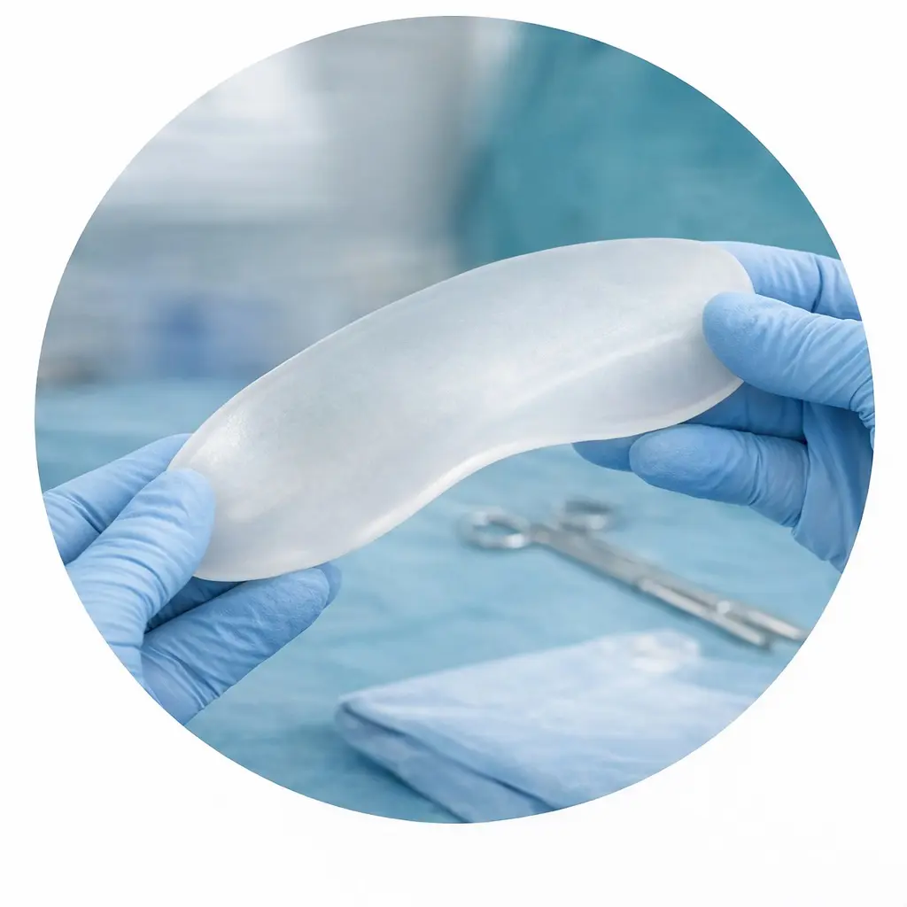
You don’t have to — that is what the assessment is for. The right operation depends on the shape, severity and stiffness of the deformity, your symptoms, and what you want to achieve. Because all of these options are available within the same practice, the recommendation is based on your chest rather than on a single preferred technique. A custom implant, for example, may suit a patient seeking purely cosmetic correction without bar surgery.
Usually, yes. Revision chest wall surgery is a specific interest of Mr Hunt’s practice — including recurrence after previous Nuss or Ravitch repair, displaced bars, and unsatisfactory results from surgery performed elsewhere. Revision cases need particularly careful imaging and planning, and often a hybrid approach, all of which is discussed openly at consultation.
Ideally yes, but it is not essential — you can refer yourself for a private consultation. A GP referral is helpful because an apparent funnel chest can occasionally be linked to another condition worth checking first, and some private insurers require a GP referral before they will authorise the consultation. If you are using insurance, check your policy.
Typically a review at around 4 weeks and again at about 12 weeks, then an annual check until the bars are removed. Most patients can travel, including by air, after the 4-week review, and are back to normal daily life and most non-contact activity by the 12-week review, with contact, collision and heavy-impact activities agreed individually.
Mr Hunt has performed pectus procedures since 2009, with a practice focused on adult and adolescent pectus deformity and complex chest wall reconstruction. The Nuss procedure itself has more than 40 years of published international evidence. Operative outcomes are recorded and reviewed, and a full discussion of results and experience is available at consultation.
Find out if surgery is right for you
Suitability for the Nuss procedure or hybrid surgery is determined at consultation. Send a brief description and photographs — Mr Hunt will review at no charge — or book directly.