Modified Ravitch
Operation
The modified Ravitch is an open operation that reshapes the chest wall directly — remodelling the abnormal costal cartilages and repositioning the breastbone. It is the main surgical option for pectus carinatum (a protruding chest) that is not suited to bracing — typically older patients with stiffer, less flexible chests — and for more complex pectus excavatum, pectus arcuatum, severely asymmetric chests and older patients with rigid chests.
Surgery at the Pectus Clinic is consultant-delivered throughout. Mr Ian Hunt assesses every patient personally and tailors the operation to the individual chest.
The modified Ravitch operation
The modified Ravitch is an open chest-wall operation. Through a single incision across the front of the chest, the abnormal costal cartilages are remodelled and the breastbone repositioned — correcting the shape of the chest directly rather than from within.
It is the principal surgical treatment for pectus carinatum (a protruding “pigeon” chest) and pectus arcuatum, particularly in older patients whose chests are stiffer and less flexible, where a brace is unlikely to work.
It is also important in pectus excavatum — for more complex, mixed or severely asymmetric chests, and in older patients with rigid chests, where a Nuss or hybrid keyhole repair may not be the right fit. It is also used for re-do (revision) surgery after a previous repair.
Mark Ravitch — origin of the operation
The open repair was first described by the American surgeon Mark Ravitch in 1949. The original operation was extensive, removing large amounts of cartilage. Over the following decades it has been refined through many variations into the modern, cartilage-sparing “modified Ravitch” used today — preserving the cartilage lining, limiting how much is removed, and adding a sternal osteotomy with temporary support where needed.
At a glance
What the modified Ravitch involves
The operation was first described by Mark Ravitch in 1949. The modified version used today is far less radical — it preserves the lining of the cartilage so the chest wall regrows in its corrected shape, and uses a sternal osteotomy and, where needed, a temporary support to hold the result.
Reshape the cartilages
The over-grown or abnormal costal cartilages driving the deformity are removed or trimmed within their lining (the perichondrium is preserved), so new, correctly-shaped cartilage regrows.
Reposition the breastbone
A controlled cut in the sternum (an osteotomy) lets the breastbone be moved into a corrected position — flattening a protrusion in carinatum, or elevating it in excavatum — and fixed there.
Support the new shape
The corrected chest is held while it heals. Increasingly this is done with low-profile titanium plates screwed to the cartilage and sternum — used for both carinatum and excavatum — designed to remain in place long term and are not removed routinely. In excavatum the correction is often combined with a bar that lifts the breastbone (a hybrid approach), removed at a later, smaller operation.
Modified Ravitch for pectus carinatum
Pectus carinatum — where the breastbone and cartilages push the chest outward — is usually treated without surgery, with a custom compression brace. Bracing works best in younger patients whose chest is still flexible.
Surgery is considered when bracing is unlikely to work or has not worked — typically a skeletally mature, rigid chest; a severe or asymmetric protrusion; an adult presenting later in life; or a patient who cannot tolerate or commit to a long bracing programme.
For these patients the modified Ravitch offers a direct surgical correction: it flattens the protrusion directly and aims to provide a durable, reliable result in carefully selected patients. It is planned individually, balancing the strength of the correction against the trade-off of an open-surgery scar.
Excavatum, mixed & complex deformity
Although carinatum is its main use, the modified Ravitch remains an important option across the range of chest-wall deformity — particularly where a keyhole (Nuss) repair is not the best fit.
It is used for markedly asymmetric or mixed excavatum–carinatum chests, for pectus arcuatum (an angulated, protruding upper sternum), for very rigid adult chests, and for revision after a previous Nuss or Ravitch that has recurred or given an unsatisfactory result.
Because Mr Hunt performs the full range of chest-wall operations — Nuss, hybrid, modified Ravitch and custom implant — the recommendation is based on the individual chest, not on a single preferred technique.
Who is suitable?
Suitability depends on the type and severity of the deformity, skeletal maturity, previous treatment, and the patient’s goals. Surgery is a positive choice, made after a full assessment by Mr Hunt.
✓ The modified Ravitch is considered for
- Pectus carinatum in a skeletally mature or rigid chest, where bracing is unlikely to work
- Severe or markedly asymmetric carinatum
- Carinatum where bracing has been declined, not tolerated, or unsuccessful
- Complex or mixed excavatum–carinatum deformity
- Pectus arcuatum and rigid adult chests
- Revision after a previous Nuss or Ravitch repair
✗ Less likely to be recommended when
- Carinatum in a younger, flexible chest — bracing is usually tried first
- A straightforward symmetric excavatum better suited to a Nuss procedure
- A more complex or asymmetric excavatum that may be better suited to a hybrid (Nuss with a mini-Ravitch) approach
- The concern is purely the appearance and a less invasive option (such as a custom implant) would suffice
- Significant medical comorbidity makes major surgery inadvisable
Assessment and planning
Every surgical patient follows a structured assessment — to confirm that an open operation is the right choice, and to plan exactly how the correction will be made.
Medical photography. Standardised clinical photographs record the starting shape and allow objective comparison after surgery.
Imaging. A CT scan of the chest with 3D reconstruction defines the pattern, severity and symmetry of the deformity and the cartilages involved, and guides the operative plan.
Heart & lung tests where indicated. In excavatum or where symptoms suggest it, an echocardiogram, cardiac MRI or lung-function/CPET testing documents any functional effect.
Anaesthetic review. Every patient is assessed before admission by a consultant anaesthetist experienced in chest-wall surgery, with the pain-management plan agreed in advance.
Planning checklist
NHS funding criteria for pectus surgery
NHS funding applies only to severe pectus excavatum (including mixed or complex deformities with a significant excavatum component). It does not cover pectus carinatum, which is treated as a self-pay procedure.
Since 2023, NHS England funds surgery for pectus excavatum only where there are very severe, objectively documented physiological symptoms. A referred patient is generally considered when there is:
- a Haller index above ~3.25 (a severe depression measured on CT or MRI), together with
- objective evidence of a functional effect — for example compression or displacement of the heart on cardiac MRI, a cardiac arrhythmia or other cardiac symptoms, significant breathlessness or exercise limitation, or swallowing difficulty (dysphagia).
Every referral is reviewed independently by a National Pectus MDT (a multidisciplinary team meeting — a panel of different specialists who consider each case together), which decides whether a patient qualifies. Those accepted are treated at one of the national pectus centres. Pectus carinatum and purely cosmetic concerns are not funded.
Mr Hunt can advise whether you are likely to meet these criteria and help arrange the appropriate referral.
The operation
The modified Ravitch is performed under general anaesthetic. The stages below describe a typical correction; the exact steps are tailored to the deformity.
Anaesthetic & preparation
You are anaesthetised, usually with a tube that allows one lung to be rested during surgery, and a regional anaesthetic block and antibiotics are given. The chest is marked, with the incision planned discreetly along the line of the chest.
Incision & exposure
A single incision is made over the front of the chest. The chest muscles (pectoralis) are gently lifted off the deformity on both sides to expose the costal cartilages, without detaching the muscle itself.
Reshaping the cartilages
The abnormal costal cartilages — typically from the 2nd to the 7th ribs — are divided and trimmed within their preserved lining on both sides, with the longer or more prominent side reduced to balance an asymmetric chest, so new cartilage regrows in the corrected shape.
Sternal osteotomy
A controlled partial cut across the breastbone lets it be brought into a corrected position — flattening a carinatum protrusion or elevating an excavatum.
Plate fixation or pectus bar
How the corrected chest is held depends on the deformity. For carinatum and most reshaping, low-profile titanium plates are shaped and screwed across the cartilages and sternum, with strong tape where helpful, and designed to remain in place long term. For excavatum, a pectus bar is passed behind the breastbone to lift it forward into the corrected position — the same principle as the Nuss and hybrid repairs — and is removed at a later, smaller operation. Some chests need a combination of both.
Cryotherapy for pain relief
The nerves running along the ribs are treated with cold (cryotherapy) on both sides. This quietens them temporarily and markedly reduces pain in the weeks after surgery; normal sensation returns as the nerves recover.
Drains, closure & recovery
One or two fine drains are placed, local anaesthetic is infiltrated, and the muscle and skin are closed in layers with dissolving stitches under the skin and a simple dressing. A chest X-ray confirms the result, and most patients are up and walking the next day.
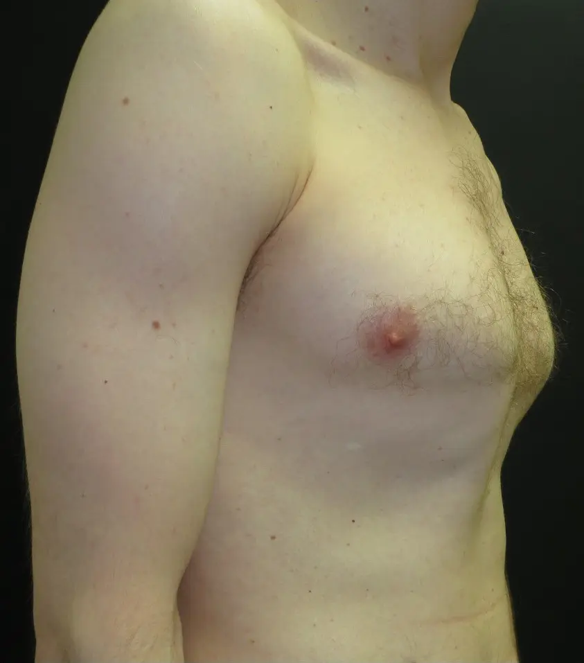
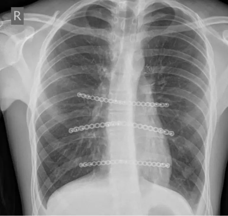
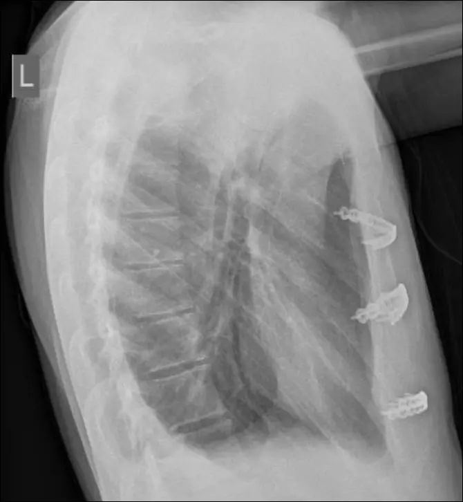
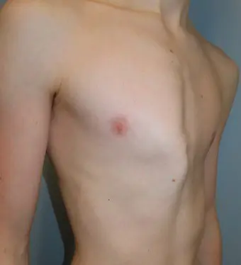
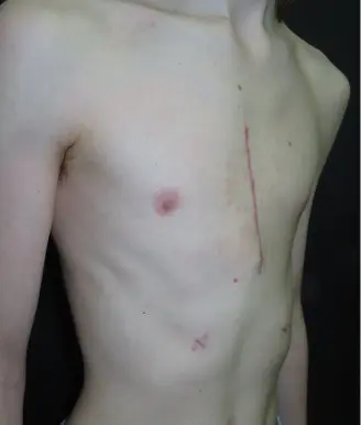
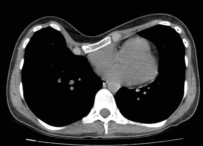
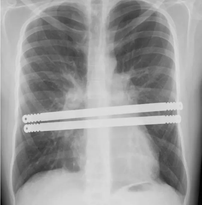
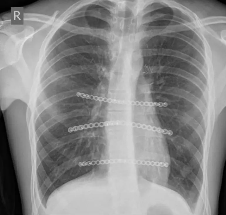
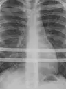
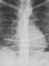
Open correction of an asymmetric pectus carinatum
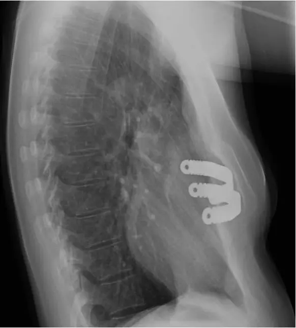
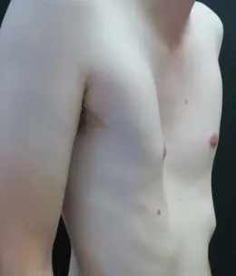
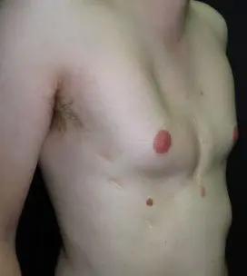
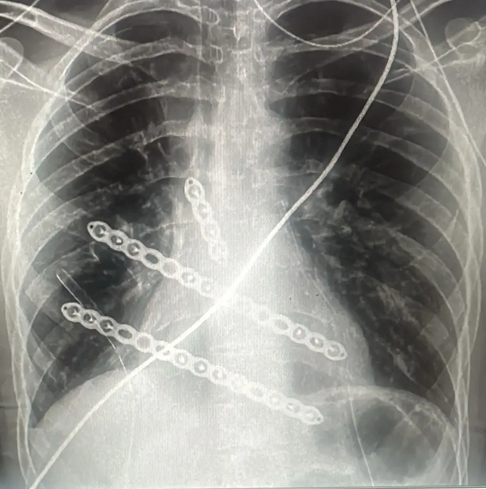
A young man with a significant, asymmetric pectus carinatum that was not suited to bracing. He was corrected with a modified Ravitch — reshaping the costal cartilages, a sternal osteotomy, and fixation with titanium plates. The images below are from his own scans and X-rays, shown with his consent.
The modified Ravitch on video
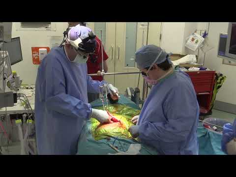
This narrated operative video, produced by Mr Hunt’s colleague Mr Joel Dunning, shows a modern modified Ravitch repair. The abnormal costal cartilages are reshaped within their lining and the breastbone repositioned and supported — the same cartilage-sparing principles used at the Pectus Clinic.
FeaturedA modified Ravitch repair for pectus — narrated operative video →
Visit Mr Dunning’s channel →This is an external video hosted on YouTube and contains surgical footage.
Recovery after a modified Ravitch
Recovery from open chest-wall surgery is steady and staged, taking about three months overall. Cryotherapy to the rib nerves during the operation helps reduce pain in the early recovery period, so most patients are home within a few days. Individual guidance is given at each review.
Typically 3–4 nights
A chest X-ray after surgery confirms the lungs are fully expanded. Pain is managed with a patient-controlled (PCA) pump and regular medication, helped by the cryotherapy. You are up and walking, with breathing exercises, the day after surgery. A soft drain is removed as it settles, and discharge follows once you are comfortable on oral painkillers — sometimes after just 2–3 days.
At home — the first 12 weeks
Settle & protect the repair
- Sleep on your back; keep good posture and bend at the hip — avoid sudden bending or twisting of the upper body.
- No heavy lifting, straining or backpacks. Walking and deep-breathing exercises are encouraged from day one.
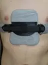
- A support band is usually worn over the wound for the first 2 weeks to limit fluid (seroma) collecting.
- Regular painkillers for about 4–6 weeks; the chest often feels numb for several months after cryotherapy. Most return to desk-based work or school by 4–6 weeks.
- Dressings are managed as advised; any soft drain is removed around 1–2 weeks. Stitches are internal — usually none to remove.
Back towards normal
- Graded return to gentle aerobic exercise — jogging, cycling, swimming — from around 6–8 weeks.
- Continue to avoid heavy lifting, weight training, backpacks and contact sport until about 3 months.
- Painkillers usually needed only occasionally.
Full activity
- Most patients return to full activity and sport by around 3 months.
- Vigorous exercise and contact or collision sports (rugby, martial arts) are left up to 6 months and agreed individually.
- Plates and screws stay in place; where a pectus bar was used it is removed at a later, smaller operation.
Contact the team — or attend A&E if urgent — if you develop: a fever; a persistent cough; worsening chest pain, especially on deep breaths; sudden breathing difficulty; redness, swelling or discharge from the wound; any injury to the chest; or a sudden change in the shape of your chest with pain.
Metalwork and bar removal
The titanium plates and screws used in a modified Ravitch are designed to remain in place long term and are not removed routinely. They hold the corrected chest while it heals and then become a stable part of the rebuilt chest wall.
Metalwork is removed only if it causes problems — for example persistent pain, a plate or screw that becomes prominent or irritating under the skin, or other issues such as loosening or infection. When removal is needed it is a much smaller procedure than the original repair: performed under general anaesthetic through the original incision, usually as a day case or with a single overnight stay, with recovery measured in days rather than weeks.
Where a pectus bar has been used — for an excavatum or a hybrid correction — this is temporary and is always removed at a planned second operation, usually after around 12 to 18 months once the chest wall has consolidated. (A bar used in a Nuss or hybrid repair stays longer — around three to four years.) Mr Hunt also removes plates, screws or bars for patients whose original surgery was carried out elsewhere, including overseas, and metalwork that has become symptomatic or displaced.
Risks and complications
The modified Ravitch is a major open operation with a good safety record in specialist hands. The main complications are grouped below by how often they occur; all are discussed openly at the consent consultation.
Exact risks vary according to deformity type, age, previous surgery and the complexity of the repair.
Common · usually minor
- Pain in the chest, controlled with painkillers and settling over a few weeks
- Numbness of the chest around the scar — usual after open surgery; often improves over months, a small area may persist
- Bruising and swelling; temporary tiredness and reduced appetite
- A visible scar across the chest (see the FAQs)
Uncommon
- Seroma (fluid) or haematoma (bleeding) collecting under the wound — occasionally needing drainage
- Wound infection — usually settles with dressings and antibiotics
- Air or fluid around the lung (pneumothorax or pleural effusion); chest infection
- Scar that heals less neatly than hoped (wide, thickened or, rarely, keloid)
Rare but serious
- Recurrence or over-correction of the deformity, occasionally needing revision surgery
- Problems with sternal healing or instability of the breastbone
- Bleeding from the internal mammary vessels requiring further surgery
- A serious anaesthetic, cardiac or respiratory complication — very rare in fit patients
Results of the modified Ravitch
In well-selected patients the modified Ravitch aims to provide a durable correction of even severe or complex deformities where less invasive routes are not suitable.
Before & after
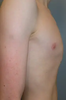
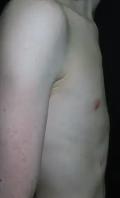
A selection of Mr Hunt’s modified-Ravitch corrections, shared with patient consent. Faces are not shown and outcomes vary between individuals.
 Before
 After
After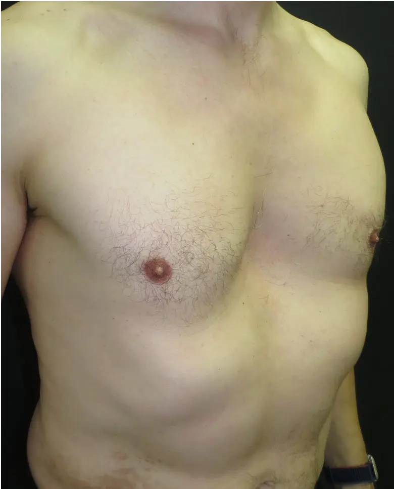 Before
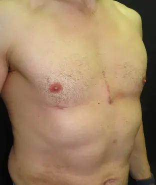 After
After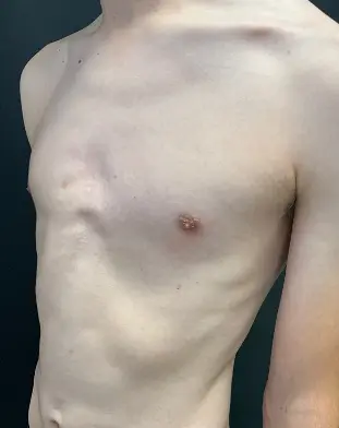 Before
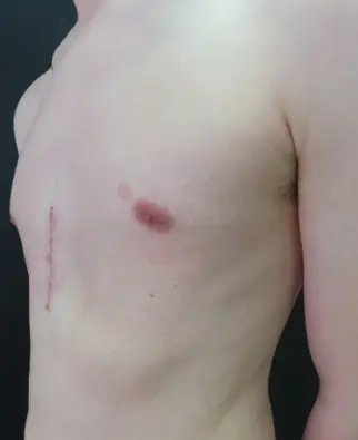 After
After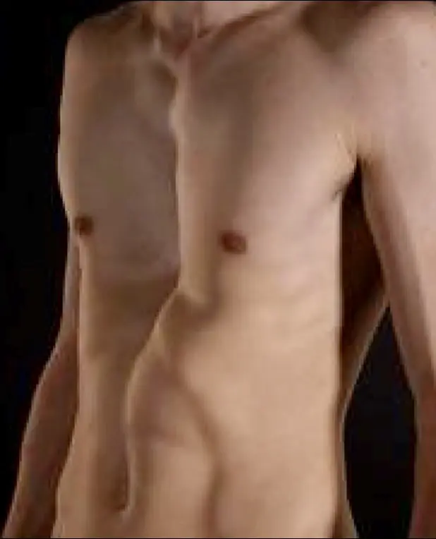 Before
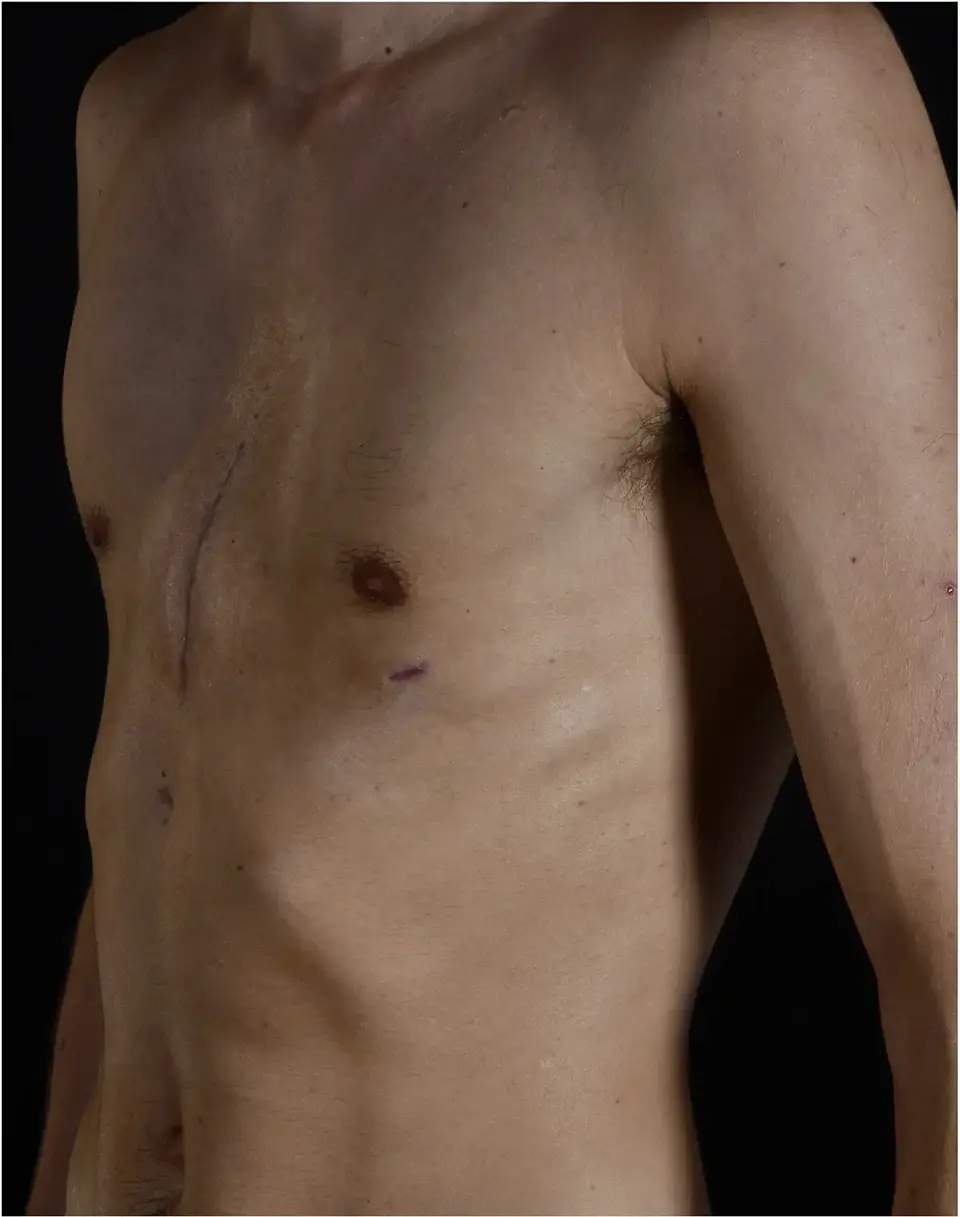 After
After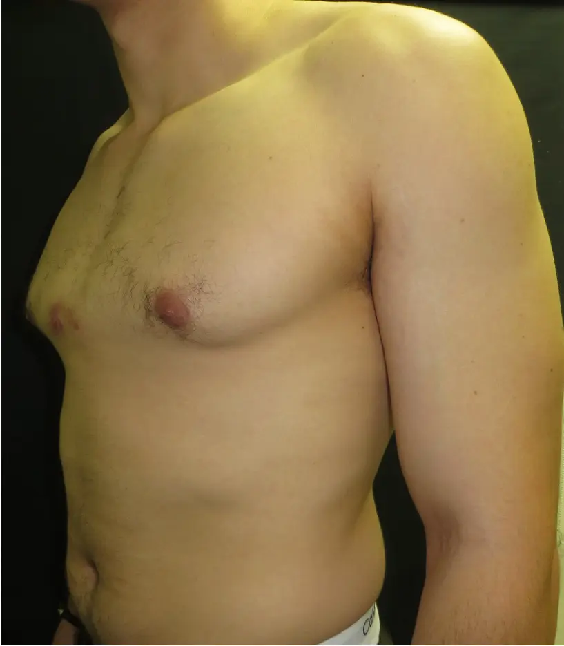 Before
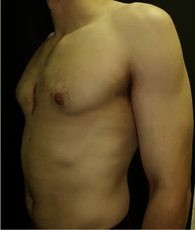 After
After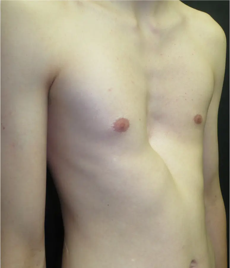 Before
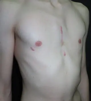 After
After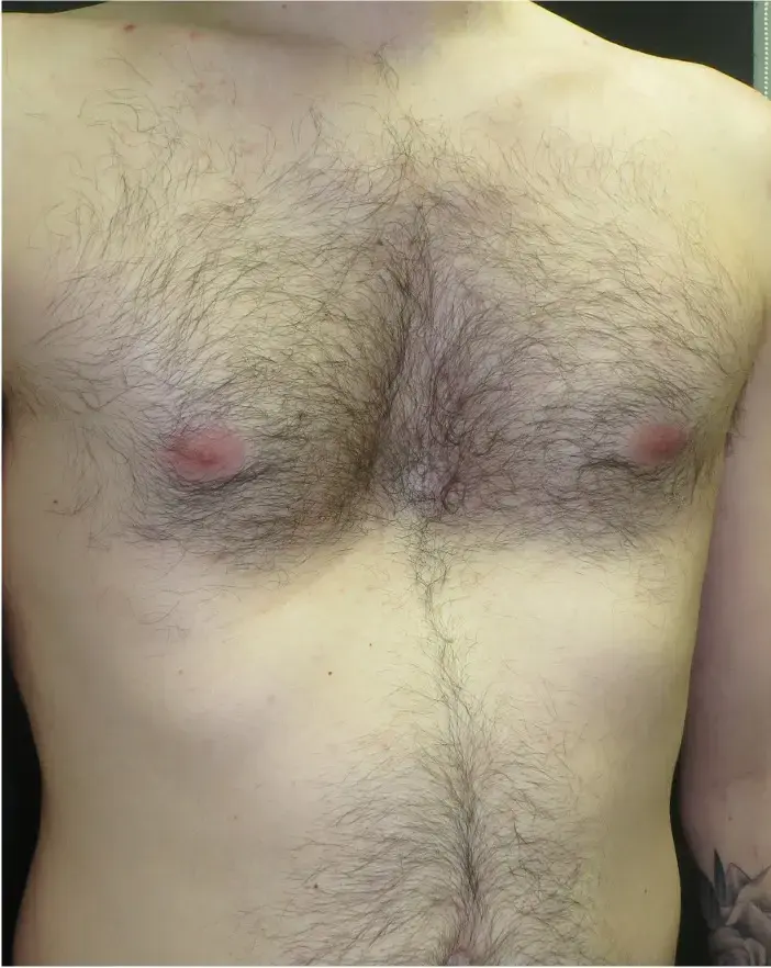 Before
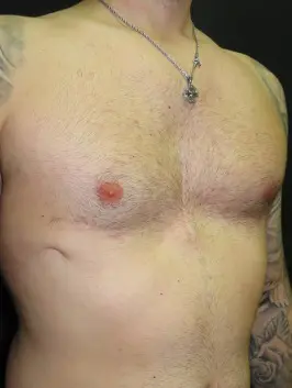 After
After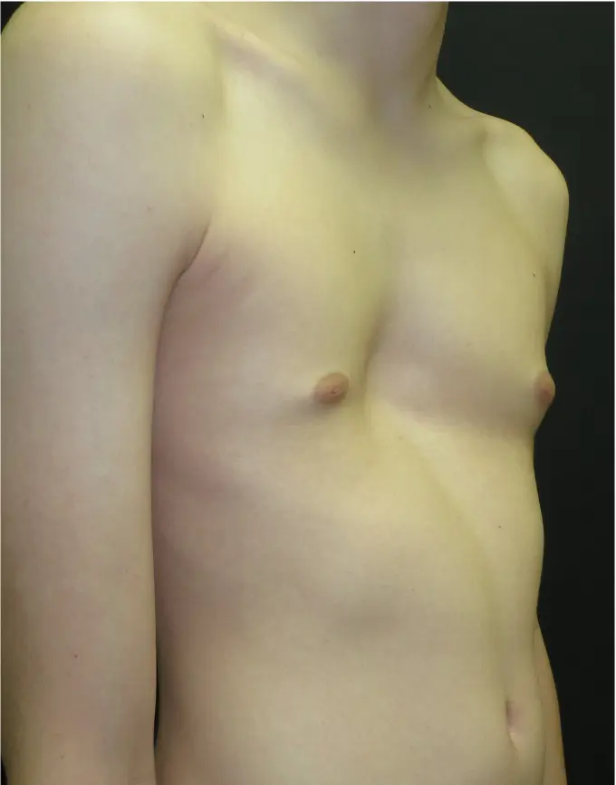 Before
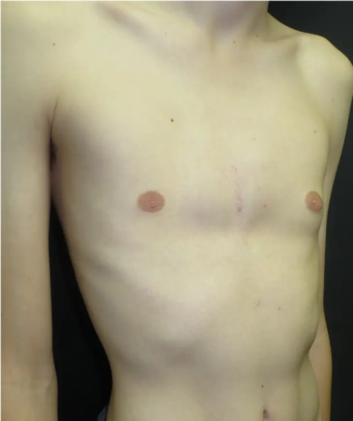 After
After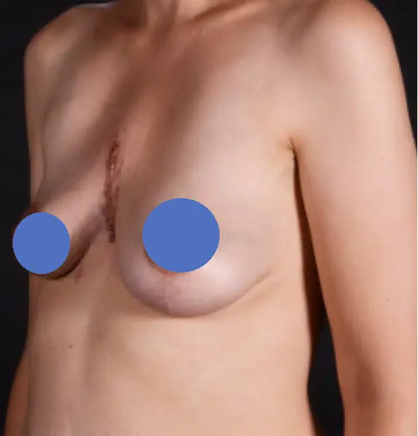 Before
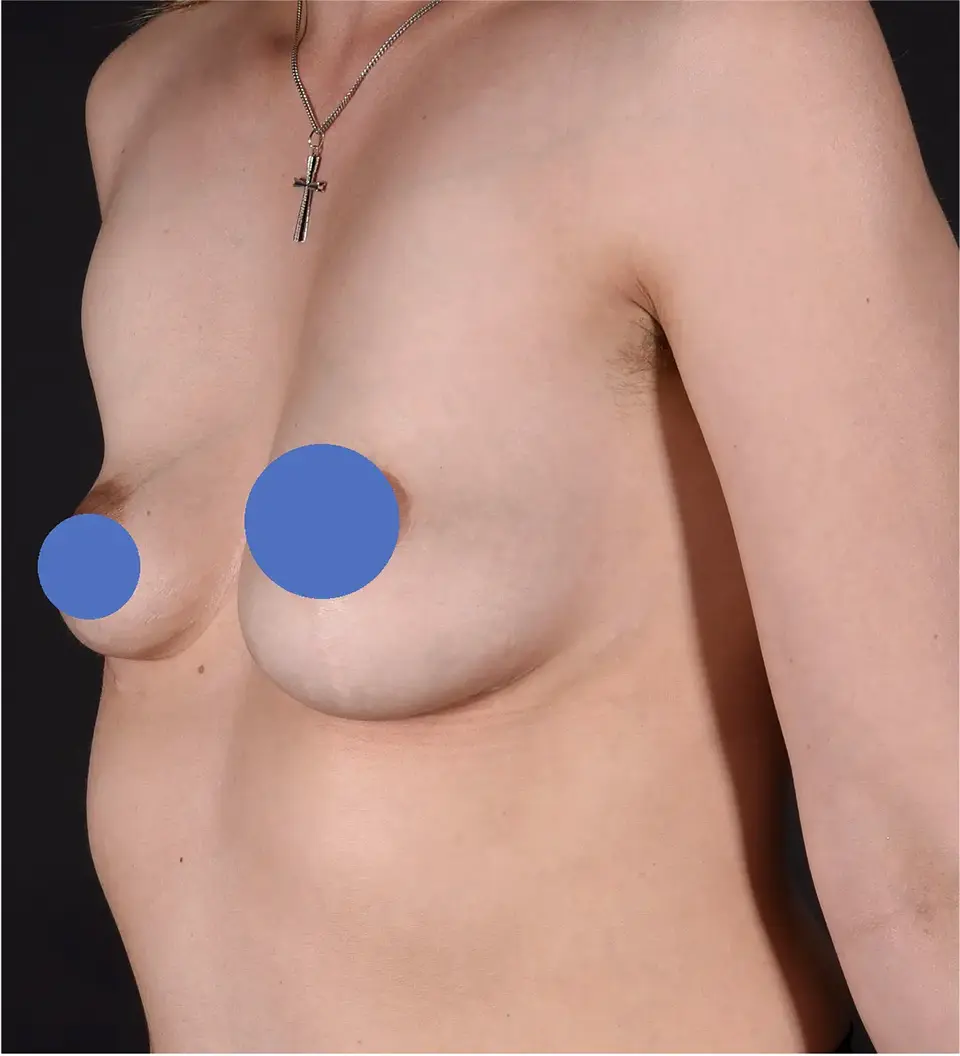 After
AfterCosts and funding for pectus surgery
How a modified Ravitch is funded depends on the deformity. Pectus carinatum is treated as a contour problem and is almost always self-pay. Severe pectus excavatum (including mixed or complex deformities with a significant excavatum component) may be funded through private medical insurance or, in defined cases, the national NHS pectus programme. The position is explained clearly at consultation so there are no surprises.
Only for severe pectus excavatum, and through a defined national pathway — pectus carinatum is not NHS-funded. For excavatum, NHS England funds surgery for patients with very severe, objectively documented physiological symptoms. The route is: referral → specialist assessment → investigations to demonstrate a genuine functional (heart or lung) effect → independent review of every case at a National Pectus MDT → if accepted, surgery at one of the national pectus centres.
The specific criteria are set out in the NHS funding criteria section above. Where an NHS route is realistic, this is discussed honestly at consultation, including the evidence a referral needs.
Private medical insurance. For pectus excavatum and complex cases, many insurers fund surgery where there is documented functional impact — symptoms plus objective findings on imaging, echocardiography or lung function testing. The assessment pathway at the Pectus Clinic is designed to provide exactly this evidence, and the team is experienced in supporting pre-authorisation applications. Carinatum is treated as a contour problem and is not usually covered.
Self-pay. For self-funding patients — including most carinatum corrections — a clear written quotation is provided after consultation, covering the surgical, anaesthetic and hospital fees and follow-up. As a guide, a surgical package is approximately £20,000, which compares well with other leading UK and international pectus centres. The package includes all hospital, surgical and anaesthetic fees, and covers every follow-up appointment and any tests or X-rays needed in the first year. Where a pectus bar is used, its later removal is quoted separately.
Modified Ravitch — FAQs
No. The modern, modified technique is much less radical: the lining of the cartilage is preserved so the chest wall regrows in its corrected shape, less cartilage is removed, and a sternal osteotomy (with internal fixation) holds the result. This gives a stronger chest wall and an easier recovery than the original 1950s operation.
Most carinatum is treated successfully with a custom brace, especially in younger patients with a flexible chest. Surgery is considered only when bracing is unlikely to work or has not worked — for example a stiff, mature chest, a severe deformity, or where bracing cannot be tolerated. The right path is decided at consultation.
Yes — it is used for selected excavatum, especially complex, asymmetric or mixed deformities, very rigid adult chests, and revision after a previous repair. For a straightforward symmetric excavatum a Nuss or hybrid repair is often preferred. The best option is decided at consultation.
Once you are asleep the operation usually takes around three to four hours. The cartilages are reshaped, the breastbone repositioned and held with titanium plates (and a bar where needed), and the rib nerves are treated with cryotherapy for pain relief.
Pain is taken seriously at every stage. During surgery the intercostal nerves are frozen (cryotherapy), which markedly reduces the need for strong painkillers afterwards. For the first day or two a patient-controlled (PCA) pump is used, then regular oral painkillers for about 4–6 weeks, reducing to occasional over-the-counter painkillers. The chest often feels numb for several months while the nerves recover.
The operation is done through a single incision across the front of the chest (occasionally extending under the breast area), positioned as discreetly as possible. Unlike the keyhole Nuss procedure it does leave a scar on the front of the chest — the main trade-off of open surgery, discussed in detail beforehand. Scars are pink and firm at first and fade over the following months.
Most patients stay around 3–4 nights (sometimes 2–3 with cryotherapy). You can usually return to desk-based work or school in 4–6 weeks, gentle aerobic exercise from about 6–8 weeks, and most sport by around 3 months. Heavy lifting, backpacks and contact sport are left until about 3 months, and vigorous or contact sport up to 6 months.
The titanium plates and screws are designed to remain in place long term and are not removed routinely — only if they cause problems such as persistent pain, prominence under the skin, loosening or infection. Where a pectus bar has been used (for an excavatum or hybrid correction) it is temporary and is removed at a later, smaller operation, usually after around 12–18 months (a bar used in a Nuss or hybrid repair stays longer, around three to four years).
In well-selected patients the correction is durable, because the cartilage regrows in its new shape and the breastbone is fixed in position. Recurrence is uncommon — under about 10% — and is more likely if metalwork is removed too early or in younger patients who are still growing. Where it occurs it is usually minor.
Metal plates or a bar may occasionally trigger airport metal detectors. There is no implant card, but you can use your clinic letter; many patients also find it helpful to keep a photo of a chest X-ray showing the metalwork on their phone. If requested, a letter for travel can be generated.
Yes, in almost all cases. Modern titanium pectus plates and bars are “MR conditional” — an MRI can be performed safely once you have recovered, provided the scan follows the manufacturer’s specified conditions. It is not automatically cleared, so always tell the radiology team the type and manufacturer of your implant beforehand; you are given details to pass on. Metalwork near the chest can cause some image artefact, though titanium produces far less than older stainless-steel devices.
The operation is generally safe, but as with any major surgery there are risks. These include bleeding (up to ~5%), air or fluid around the lung (pneumothorax, or pleural effusion in under 1%), wound infection (deep infection involving the metalwork is rare), a fluid collection under the skin (seroma, managed with a drain and support band), temporary numbness around the scar, and displacement of a plate or bar (around 2%, mostly in the first six weeks). Persistent pain or an unstable sternum are uncommon, and injury to the heart or major vessels is extremely rare. Risks are discussed fully before surgery.
Surgery for pectus carinatum is treated as a contour procedure and is generally self-pay (not NHS-funded). Correction of severe pectus excavatum with documented functional symptoms may be covered by private insurance, or meet the criteria of the national NHS pectus pathway. Your situation is explained clearly at consultation.
Find out if the modified Ravitch is right for you
Suitability is determined at consultation. Send a brief description and photographs — Mr Hunt will review — or book directly.