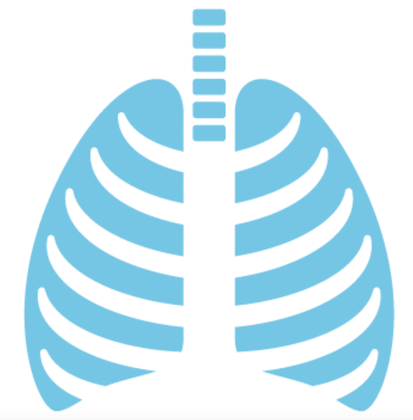
Posture & Pectus
Chest wall deformities frequently generate secondary postural changes that develop gradually alongside the underlying condition. Understanding these changes — and where in the body they arise — is an important part of assessment and management.
Why pectus causes postural problems
The chest wall is structurally central to the body. When its shape is abnormal, the surrounding musculoskeletal system adapts — often in ways that compound progressively over time.
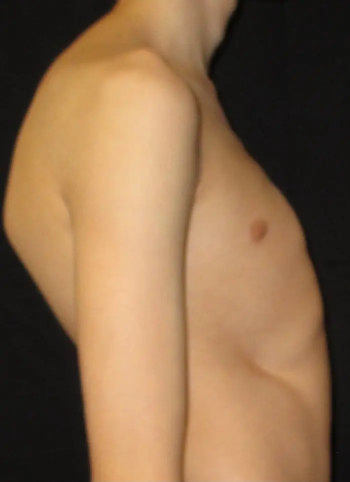
In pectus excavatum, the depressed sternum and restricted thoracic expansion drive a characteristic pattern of thoracic kyphosis, rounded shoulders and forward head displacement. In pectus carinatum, a similar but distinct pattern emerges — often with prominent upper thoracic extension and associated shoulder tension. In mixed or asymmetric deformities, the picture is more variable and typically includes lateral imbalance.
These changes matter clinically. They affect breathing mechanics, contribute to discomfort, and can progressively entrench if not addressed during adolescent growth. Many are also amenable to improvement through targeted physiotherapy.
At a glance
Forward head posture
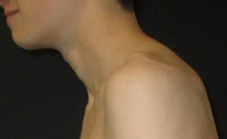
Forward head posture is one of the most consistently observed secondary changes in pectus patients. As the upper thoracic spine flexes into kyphosis, the head shifts forward relative to the shoulders to maintain a level gaze.
For every inch the head moves forward, the effective weight borne by the cervical spine increases substantially — this creates chronic tension in the posterior neck muscles and upper trapezius, which are frequently tender in pectus patients even without a specific diagnosis of neck pain.
In pectus carinatum, a similar pattern can develop through habitual shoulder rounding, though the degree of forward head translation tends to be less pronounced than in PE. In younger patients, these changes are largely soft-tissue driven and more amenable to postural correction.
How to recognise it
- Head positioned noticeably in front of the shoulders on lateral view
- Chin protrusion and loss of the natural cervical curve
- Tightness or tenderness in the back of the neck and upper trapezius
- Upper cervical prominence at the base of the skull
- Almost always associated with rounded upper back and elevated shoulders
Thoracic kyphosis
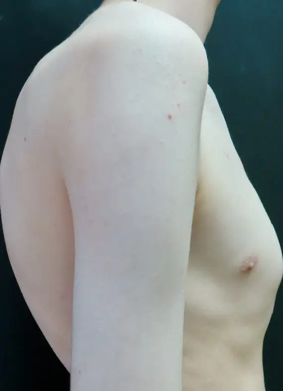
Thoracic kyphosis — excessive rounding of the upper and mid-back — is the most consistent and clinically significant postural change associated with pectus deformity, particularly pectus excavatum.
The sunken anterior chest wall effectively creates a void that the spine accommodates by flexing forward. Over time, the thoracic extensor muscles lengthen and weaken while the anterior chest and pectoral structures shorten, creating a self-reinforcing cycle that is progressive if unchecked.
The degree of kyphosis does not always correlate directly with the severity of the pectus deformity. Some patients with mild PE develop significant kyphosis; others with deep excavatum maintain a relatively straight thoracic spine. The relationship is influenced by age, growth, muscle tone and habitual posture.
How to recognise it
- Visibly rounded upper and mid-back on lateral view
- Reduced thoracic extension range when asked to stand tall
- Tightness across the upper back and between the shoulder blades
- Forward head posture developing secondarily
- Reduced thoracic rotation and lateral flexion
- May be associated with mild scoliotic curvature in asymmetric presentations
Rounded & protracted shoulders
Rounded, protracted shoulders are a near-universal finding in pectus patients. As the thoracic spine flexes and the chest wall changes shape, the pectoral muscles shorten adaptively and the scapulae rotate forward on the thorax.
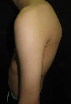
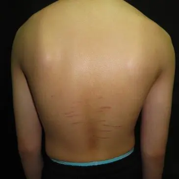
note development of lower back stretch marks
Stretch marks (striae) over the shoulders and upper back are a recognised finding in rapidly growing adolescents with pectus deformity. They reflect accelerated growth and skin tension during the period of greatest chest wall change, and are a useful clinical marker of recent or active growth.
This anterior tipping of the shoulder complex reduces the subacromial space and compresses the glenohumeral joint — contributing to shoulder discomfort and reduced overhead range of motion that is commonly reported alongside the chest wall deformity. It also drives scapular protraction and inhibits the lower and middle trapezius.
How to recognise it
- Shoulders rolled forward at rest — visible on both lateral and frontal views
- Reduced space between chin and shoulders — neck appears shortened
- Tightness across the anterior chest and pectoral muscles
- Difficulty sustaining a retracted shoulder position without active effort
- Associated upper back tightness and fatigue with sustained upright posture
Scapular winging
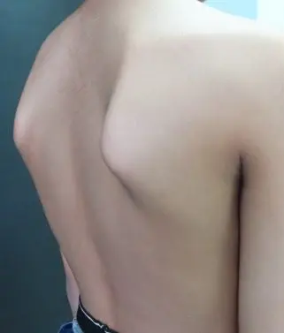
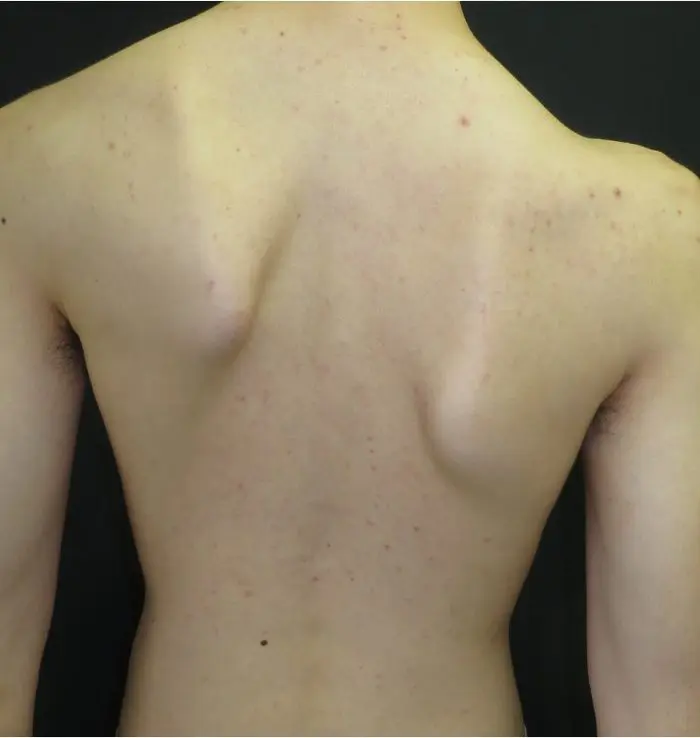
Scapular winging — where the medial border or inferior angle of one or both scapulae lifts away from the thorax — is a specific manifestation of scapular instability seen in a proportion of pectus patients.
It arises from weakness or inhibition of the serratus anterior and lower trapezius, which are responsible for holding the scapula flat against the posterior thorax. In pectus patients, the altered chest wall contour changes the mechanical advantage of these muscles, making effective scapular stabilisation more effortful.
Winging may be subtle — noticed only during push-ups or arm elevation — or pronounced enough to be visible at rest. It is not in itself painful, but it is associated with reduced shoulder function and is a marker of broader scapular control deficit.
How to recognise it
- Medial scapular border visibly prominent on posterior view
- Winging worsens or becomes visible during press-ups or wall push
- Asymmetric scapular position, often more pronounced on one side
- Associated rounded shoulder posture and reduced shoulder strength
- May be present at rest or only apparent with loading
Chest wall mechanics & breathing pattern
The altered shape of the chest wall directly restricts thoracic movement, and breathing pattern adapts to work around these restrictions — often in ways that generate their own secondary consequences.
In pectus excavatum, the reduced anterior-posterior thoracic dimension limits the diaphragm's range of excursion. The body compensates by recruiting accessory muscles in the neck and upper chest — a pattern that, when habitual, contributes to upper trapezius tension, neck discomfort and a sense of restricted breathing even at rest. Lateral rib expansion and posterior thoracic breathing are often reduced, with the lower ribs becoming progressively less mobile over time.
How to recognise it
- Visible upper chest movement during normal breathing at rest
- Reduced lateral and posterior rib expansion on deep inhalation
- Accessory muscle tension in the neck during breathing
- Paradoxical abdominal movement (abdomen draws in on inspiration)
- Breathlessness disproportionate to activity level
Uneven shoulder height
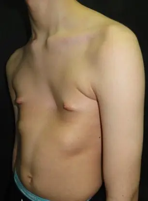
Asymmetric shoulder height is a common feature in mixed and asymmetric pectus presentations, and occasionally in patients with predominantly unilateral PE or PC. It reflects lateral trunk asymmetry that originates in the chest wall itself.
Unlike scoliosis — where uneven shoulders arise from lateral spinal curvature — in most pectus patients the underlying driver is chest wall asymmetry and associated soft-tissue imbalance rather than a primary spinal problem. However, the two can coexist, and formal assessment is warranted when shoulder asymmetry or lateral trunk tilt is present.
The degree of shoulder asymmetry seen on standing does not always reflect the severity of the underlying chest wall asymmetry directly — soft-tissue compensation and habitual posture can exaggerate or partially conceal the underlying structural difference.
How to recognise it
- Visible height difference between the two shoulders on posterior or frontal view
- Associated lateral trunk tilt on standing
- One scapula sitting higher or more laterally than the other
- Neck appearing to lean or tilt to one side at rest
- May be associated with mild scoliotic curvature — formal assessment required
Scoliosis & pectus
A mild degree of lateral spinal curvature — scoliosis — is seen in a proportion of pectus patients, particularly those with asymmetric or mixed chest wall deformity. In most cases this is mild and functional rather than structural, and does not require specific spinal treatment. However, it warrants assessment to distinguish it from primary scoliosis, which may progress independently.
Where scoliosis coexists with pectus, it can contribute to postural asymmetry, uneven rib heights and the appearance of lateral trunk tilt. The relationship between the two conditions is variable — chest wall correction does not reliably resolve associated scoliosis, and any significant curvature requires separate evaluation.
Read more about scoliosis & pectus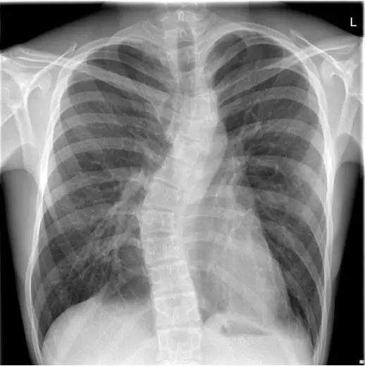
Core control, lumbar lordosis & anterior pelvic tilt
The lower end of the postural chain is often affected in pectus patients — anterior pelvic tilt, lumbar hyperlordosis and poor core control frequently accompany the changes seen higher up.
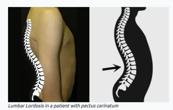
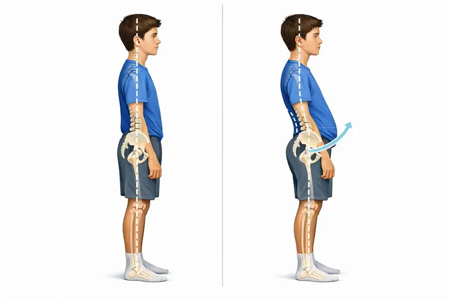
When the thoracic kyphosis is pronounced, the lumbar spine compensates by increasing its lordotic curve to maintain upright balance. This is associated with anterior pelvic tilt, in which the front of the pelvis drops and the back rises — stretching the hip flexors and compressing the posterior lumbar structures. Lower abdominal control is frequently compromised, and rib flare — already a feature of some chest wall deformities — is often worsened by the inability to maintain lower rib position against gravity.
How to recognise it
- Anterior pelvic tilt and increased lumbar lordosis on lateral view
- Lower abdominal protrusion, particularly in adolescents
- Difficulty maintaining a neutral spine during low-load exercise
- Rib flare that worsens with abdominal loading
- Inability to consciously maintain lower rib position during expiration
The combined postural picture
The individual changes described above rarely occur in isolation. In most pectus patients seeking assessment, several of these patterns are present simultaneously and interact with one another to produce a recognisable overall postural profile.
The chain runs from head to pelvis — forward head posture, thoracic kyphosis, rounded and protracted shoulders, scapular instability, altered breathing mechanics and anterior pelvic tilt tend to cluster together. In asymmetric presentations, lateral imbalance is added to this picture.
Assessment of standing posture — in multiple planes, at rest and during movement — is a standard part of every consultation at the Pectus Clinic.
The full chain — head to pelvis
- Head forward, chin protruding, reduced cervical lordosis
- Upper back rounded into thoracic kyphosis
- Shoulders protracted, elevated and internally rotated
- Scapular winging — medial borders prominent posteriorly
- Chest compressed or prominent, narrow thoracic aperture
- Lower ribs flared, rib control reduced
- Lumbar hyperlordosis and anterior pelvic tilt
- In asymmetric pectus: lateral trunk tilt and uneven shoulders
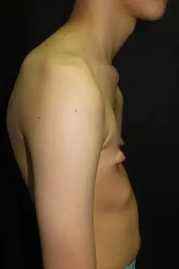
Postural assessment is part of every consultation
Every patient seen at the Pectus Clinic receives a structured assessment that includes evaluation of secondary postural changes, breathing pattern and thoracic mobility. Written exercise resources and physiotherapy referral are provided where appropriate.