
Rib Flare
Lower rib prominence
Rib flare describes the outward protrusion of the lower costal margin — the cartilaginous border formed by the lower ribs. It may be an isolated finding or occur alongside other chest wall conditions, and ranges from a minor cosmetic concern to a source of significant discomfort and functional difficulty.
“ Not every case of rib flare warrants active intervention — but when it causes real discomfort, limits exercise, or creates significant distress, it deserves careful assessment rather than dismissal. The goal is always to understand what is driving the problem before deciding whether, and how, to treat it. ”
Mr Ian Hunt — Consultant Thoracic & Chest Wall Surgeon
What is rib flare?
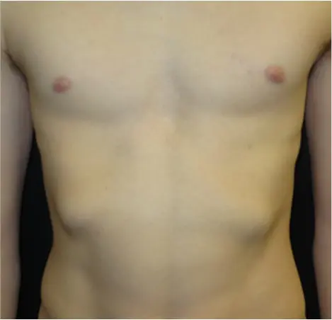
Rib flare refers to the outward protrusion or widening of the lower costal margin — the arched border formed by ribs 6 to 10 as they meet the cartilage connecting them to the sternum. In a typical chest, this border sits close to the body wall. In rib flare, it angles outward, creating a visible and often palpable shelf or ridge beneath the lower chest.
The degree of flaring varies considerably. In mild cases, the ribs protrude only slightly and may not cause any symptoms at all. In more pronounced cases, the lower ribs can project substantially beyond the line of the abdominal wall, causing discomfort during physical activity, difficulty lying on one side, and significant cosmetic concern. The condition may affect one side only (asymmetric) or both sides to varying degrees. When bilateral, the left side is more commonly the more prominent — a pattern seen frequently in clinical practice, though the reason for this asymmetry is not fully established.
Rib flare is most commonly encountered in association with pectus deformity — particularly pectus excavatum and pectus carinatum — where it frequently forms part of the overall lower chest wall abnormality. It may also occur as an isolated finding, or arise from poor posture and muscle imbalance, hypermobility of the costal cartilages, or connective tissue disorders such as Ehlers–Danlos syndrome. In some individuals, costal cartilage laxity plays a central role. Careful assessment is needed to understand which mechanism is driving the problem before treatment decisions are made.
Pain is an important and sometimes underappreciated feature. Prominent rib flare can cause intercostal discomfort, particularly during exercise, prolonged sitting, or when pressure is applied to the lower chest wall. In some patients, the relationship between rib flare and pain is more complex: laxity or instability at the lower costal margin can overlap with the clinical picture of slipping rib syndrome — a condition in which a hypermobile lower rib produces catching, clicking or sharp pain with certain movements. Where this overlap is present, accurate characterisation of the anatomy and mechanics is essential to guide treatment.
Rib flare and pain. Not all patients with rib flare have pain, but many do. Where lower costal margin pain or instability is present — particularly with a clicking, catching or reproducible quality — the possibility of an associated slipping rib syndrome component should be considered and assessed carefully.

Key Facts
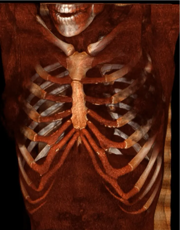
Chest CT with 3D reconstruction
This image highlights the costal cartilage at the front of the chest, demonstrating bilateral rib flare with splaying of the lower costal margin. The 9th rib tips appear not to join the costal cartilage above, as is typically described. This patient presented with pain and bilateral slipping rib syndrome.
Image used with patient consent for educational purposes. Individual anatomy varies.
The anatomy of rib flare
The lower costal margin is formed by the fused cartilages of ribs 7–10, which meet at the costal arch. In patients with rib flare, this arch is widened and the lower chest wall protrudes laterally, often more prominently on the left.
Rib flare is frequently associated with pectus deformity, and may also reflect underlying chest wall geometry, posture, or soft tissue factors. In some patients, the costal cartilage configuration contributes to pain or instability at the rib tips.
Rib swooping
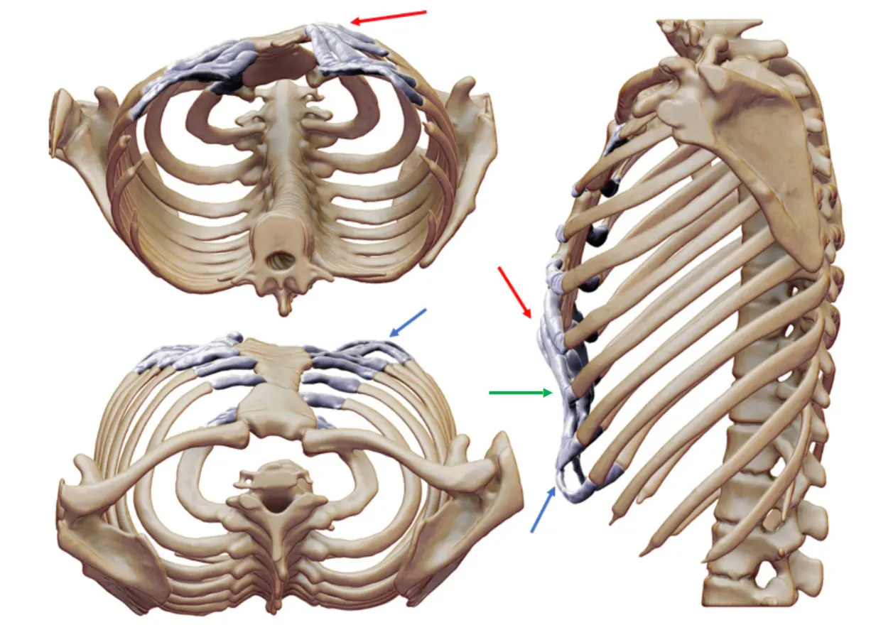
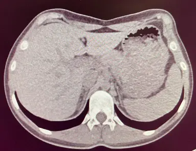
In some patients, the lower ribs and costal cartilages do not follow a smooth rounded curve from the costal arch towards the sternum. Instead, the cartilages appear to flatten or “swoop” upwards across the front of the chest, often creating a broader rib flare appearance.
This rib shape is commonly seen with pectus carinatum, but it can also occur on its own. It may contribute to the visual impression of rib flare, chest wall asymmetry, or a flattened lower anterior chest contour.
Signs and symptoms
Rib flare presents differently across patients. The symptoms that matter most — cosmetic, functional, or both — vary with severity, underlying cause and individual circumstance.
Visible protrusion and appearance
The lower ribs angle outward visibly, with the contour of the lower chest wall appearing rounded, flared, or prominent. Many patients — particularly adolescents — describe body image concerns, avoidance of swimming or sport, and self-consciousness that is taken seriously in assessment.
Asymmetry and posture
Asymmetric flare is common, more typically pronounced on the left. This may appear as a lateral imbalance or rotational component and is frequently associated with thoracic kyphosis, forward head posture, or muscle imbalances that a physiotherapy assessment can help address.
Intercostal discomfort
Pain or aching along the lower costal margin, particularly during exercise, prolonged sitting, or when the ribs are compressed. In patients with associated slipping rib syndrome, pain may be sharp or episodic.
Psychological impact
For many patients — particularly adolescents and young adults — the psychological burden of rib flare can be as significant as any physical symptom. The visible prominence of the lower chest wall affects body image at a developmentally sensitive time, with effects that may persist into adulthood.
Avoidance behaviours are common: patients describe self-consciousness about wearing fitted clothing, avoiding swimming, sport, or situations where the chest wall might be visible. Anxiety about appearance and reduced self-confidence are recognised as valid aspects of clinical assessment, independent of the objective degree of flare.
The Pectus Clinic takes psychological impact seriously. Where appropriate, referral for specialist psychological support is available alongside clinical assessment and treatment planning.
How severity and shape vary
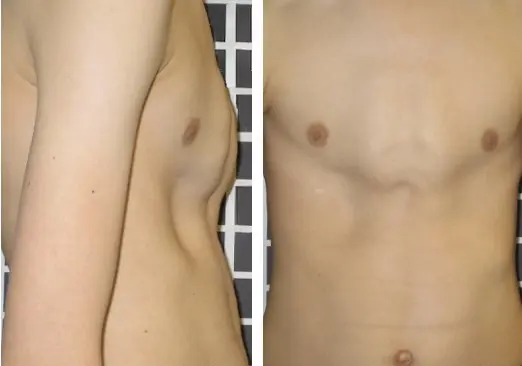
Rib flare presents across a wide spectrum. In mild cases it may be barely noticeable at rest and visible only when the arms are raised. In moderate forms the lower costal margin is clearly prominent, typically more so on the left, and the shelf-like contour of the ribs can be seen at rest. Severe cases involve marked bilateral flare, often in association with pectus deformity, with significant impact on chest wall contour and — in some patients — symptoms of discomfort or pain. No two presentations are identical.

Mild — left-sided predominance
Subtle unilateral flare, most apparent on the left. The lower costal margin is slightly widened but the contour difference is modest and often only noticeable on close inspection or when the arms are elevated.
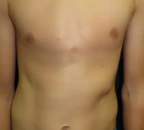
Moderate bilateral — shelf-like left costal margin
Clearly visible bilateral flare, more prominent on the left, creating a shelf-like lower costal margin. The asymmetry is apparent at rest and may be associated with mild postural changes.
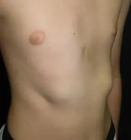
Moderate bilateral — characteristic right-sided cleft
Bilateral flare with a characteristic notch or cleft visible at the right costal margin, reflecting an anatomical variant in the way the lower costal cartilages meet. A distinct and recognisable morphological pattern.
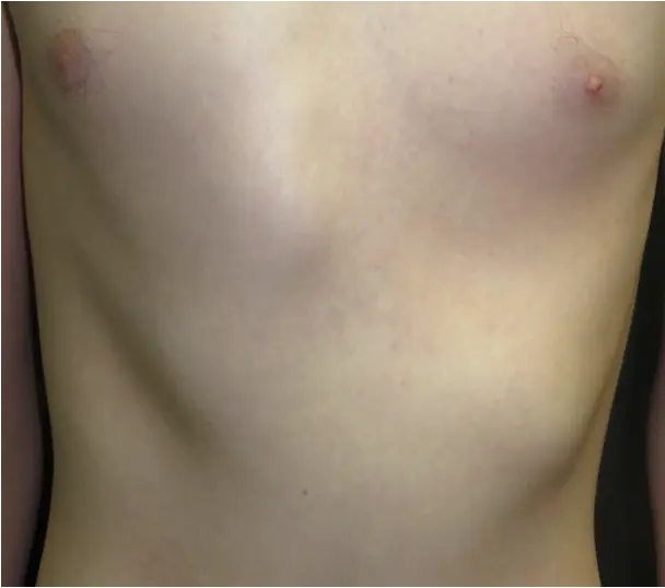
Moderate — atypical morphology
An unusual pattern of rib flare that does not conform to the typical bilateral or left-predominant presentation. Careful assessment is important as morphological variation influences treatment planning.
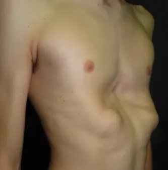
Severe bilateral — with pectus excavatum
Marked bilateral rib flare coexisting with pectus excavatum. The combination produces a significant chest wall deformity and often requires an integrated assessment and treatment approach addressing both the sternal depression and the lower costal flare.

Female — bilateral with mild pectus excavatum
Bilateral rib flare in a female patient with associated mild pectus excavatum. Rib flare in female patients may be partially obscured by breast tissue but remains clinically significant, particularly where there is associated pain, asymmetry, or deformity.
How rib flare is assessed
Rib flare is primarily a clinical diagnosis. In most patients, careful inspection, surface measurement and a structured symptom history are sufficient to characterise the deformity, assess its severity and guide treatment decisions. Photographs in standard positions are taken at baseline to document appearance and track change over time.
Assessment includes symmetry, degree of lateral flare, associated rib swooping, thoracic kyphosis and the presence of a related pectus deformity. Where costal margin tenderness, snapping or intercostal discomfort are reported, slipping rib syndrome is also considered. Physiotherapy assessment — particularly of posture, breathing pattern and muscle balance — forms part of the initial pathway for most patients.
When imaging is needed
Imaging is not routinely required. CT chest may be requested selectively — to characterise a coexisting pectus deformity, evaluate the costal cartilages in suspected slipping rib syndrome, or inform surgical planning. Where CT is performed, three-dimensional reconstruction provides detailed chest wall anatomy. Dynamic ultrasound of the costal margin may be used where slipping rib syndrome is clinically suspected.

Associated pectus excavatum
Where rib flare occurs alongside pectus excavatum, a more structured assessment is carried out — including CT chest with Haller Index calculation and, where indicated, cardiopulmonary exercise testing and cardiac evaluation.
Full assessment pathway — Pectus Excavatum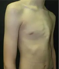
Associated pectus carinatum
Where rib flare coexists with pectus carinatum, assessment is extended to evaluate the full chest wall configuration — including the degree of sternal protrusion, overall symmetry and suitability for bracing or combined surgical correction.
Full assessment pathway — Pectus CarinatumTreating rib flare
Treatment decisions are individual and depend on severity, symptoms, underlying cause, and patient goals. Not all cases of rib flare require active intervention.
Observation & monitoring

For mild or asymptomatic rib flare, watchful waiting with regular review is appropriate. Many cases in adolescence stabilise without treatment. Monitoring includes periodic clinical assessment and photography to track any progression.
Associated pectus deformity. Where rib flare occurs alongside pectus excavatum or pectus carinatum, treatment planning extends to address the full chest wall picture. Management of the associated deformity follows its own structured pathway and is discussed separately at consultation.
Exercise & Postural Retraining
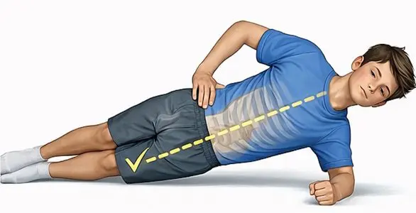
A structured programme of targeted exercises — focused on activation of the core abdominal, pelvic and lower back muscles — is often the first active step in management. Breathing retraining and postural correction can reduce the appearance of rib flare and improve associated discomfort.
Associated pectus deformity. A targeted exercise programme is also a core part of the non-surgical treatment pathway for associated pectus deformities. Where specialist input is needed, we can refer within our physiotherapy network.
External Rib Flare Bracing
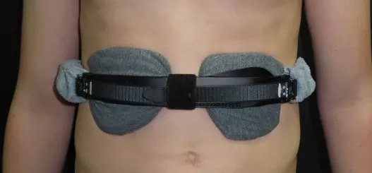
A custom-fitted brace applies directed pressure to the lower costal margin, encouraging the ribs inward over time. Bracing for rib flare is most effective during adolescence when the chest wall is still growing and flexible. It requires commitment to consistent daily wear and regular clinical review to assess progress and adjust the fit.
Associated pectus deformity. Bracing for rib flare may be combined with or run alongside bracing for an associated pectus deformity. Where both conditions are present, the treatment programme is planned together.
Rib Cartilage Resection
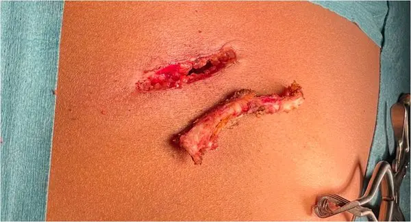
In selected patients with well-defined lower rib flare — especially if painful or associated with slipping rib syndrome — resection of a portion of the offending costal cartilage can produce meaningful improvement. It is performed under general anaesthetic with a short hospital stay.
Rib flare and pain. Not all patients with rib flare have pain, but many do. Where lower costal margin pain or instability is present — particularly with a clicking, catching or reproducible quality — the possibility of an associated slipping rib syndrome component should be considered and assessed carefully.

Rib Stabilisation & Reconstruction
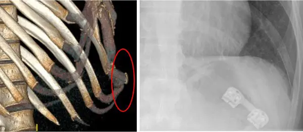
In complex cases — including those with significant asymmetry, prior rib surgery, connective tissue laxity or multilevel involvement — more extensive surgical reconstruction may be required. This may involve rib fixation techniques using absorbable or titanium hardware, or a combined approach with pectus correction where a coexisting deformity is present.
Complex chest wall problems. Rib stabilisation and reconstruction is part of a broader surgical toolkit for complex and revision chest wall cases. Where rib flare forms part of a more complex picture — including chronic costal margin pain, instability, or failed prior surgery — a full assessment at the Rib Injury Clinic is the appropriate starting point.
Combined pectus surgery
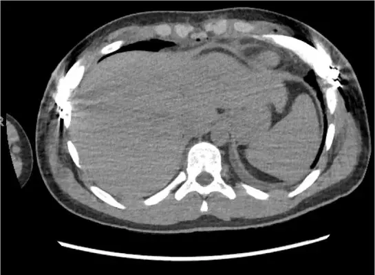
Where rib flare coexists with pectus excavatum or pectus carinatum, both conditions can often be addressed in a single operation. When performing a Nuss or hybrid procedure for pectus excavatum, associated rib flare can be corrected simultaneously using a targeted rib flare technique. When performing a modified Ravitch procedure for pectus carinatum, costal cartilage excision can be extended to address the lower costal margin at the same time. Combined surgery avoids the need for separate procedures and is planned carefully with cross-sectional imaging and a full preoperative discussion.
Associated pectus deformity. Combined surgery is discussed and planned as part of the full assessment for pectus excavatum or pectus carinatum. Full details of each surgical pathway are on the condition pages.
Frequently asked questions
Is surgery always needed for rib flare?
No. Many patients with rib flare manage well with non-surgical approaches — particularly targeted exercise, postural retraining +/- physical therapy, and in adolescents, bracing. Surgery is considered only when symptoms are significant, non-surgical treatment has been insufficient, and the expected benefit outweighs the risks. The decision is always made jointly with the patient.
Does rib flare get worse over time?
It varies. In adolescents, rib flare may worsen during growth spurts and then stabilise once skeletal maturity is reached. In adults, the bony structure is fixed, though the appearance can change with changes in body composition or posture. Regular review helps to track any progression.
Can targeted exercise alone correct rib flare?
A targeted exercise programme can improve the appearance and symptoms of rib flare, particularly when posture and muscle balance are contributing factors. It is unlikely to fully correct a structural bony or cartilaginous deformity on its own, but it is a valuable part of the management pathway and often produces meaningful benefit — including alongside bracing or as preparation for, or recovery after, surgery.
What is the recovery after surgery?
Recovery depends on the type and extent of surgery. For rib cartilage resection, most patients are discharged within one to two days and return to light activities within two to three weeks. More extensive reconstruction requires a longer recovery. Detailed expectations are discussed during the pre-operative consultation.
Will my insurance cover treatment for rib flare?
Insurance coverage varies between providers and policies. Functional or symptomatic rib flare is more likely to be covered than treatment sought purely for cosmetic reasons. The clinic's administrative team can assist with pre-authorisation queries. Further details are available on the Fees & Funding page.
Is rib flare related to my connective tissue condition?
It can be. Hypermobility and connective tissue disorders such as Ehlers–Danlos syndrome are associated with costal cartilage laxity, which can contribute to rib flare. If this is suspected, connective tissue review is coordinated before treatment decisions are made, as it affects both the assessment and the choice of approach.
Arrange a specialist assessment
All patients are seen by Mr Ian Hunt personally. Consultations include a full clinical assessment, review of any existing imaging and a clear, unhurried discussion of the options available.