
Pectus Carinatum
Pigeon Chest
The second most common chest wall deformity, caused by abnormal growth of the costal cartilages, which push the sternum outwards. It typically becomes apparent during the adolescent growth spurt and can be effectively treated with bracing or, in selected cases, surgery.
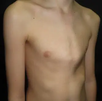
What is pectus carinatum?
Pectus carinatum occurs when the cartilages connecting the ribs to the breastbone grow too rapidly, pushing the sternum forward and creating a prominent, protruding chest wall. The term comes from the Latin carina, meaning keel — reflecting the characteristic outward appearance.
Unlike pectus excavatum, in which the sternum is depressed inwards, pectus carinatum results in a convex chest profile. It most commonly involves the lower costal cartilages and sternum, and it typically becomes noticeable during the rapid growth phase of puberty, often appearing to emerge quite suddenly between the ages of 11 and 15.
It is not present at birth in most cases. Many families notice the change progresses with height gain and then stabilises once skeletal maturity is reached. This growth-dependent behaviour is important clinically, as it means bracing treatment — which works by applying external corrective pressure — is most effective during active growth.
Condition at a Glance
The anatomy of pectus carinatum
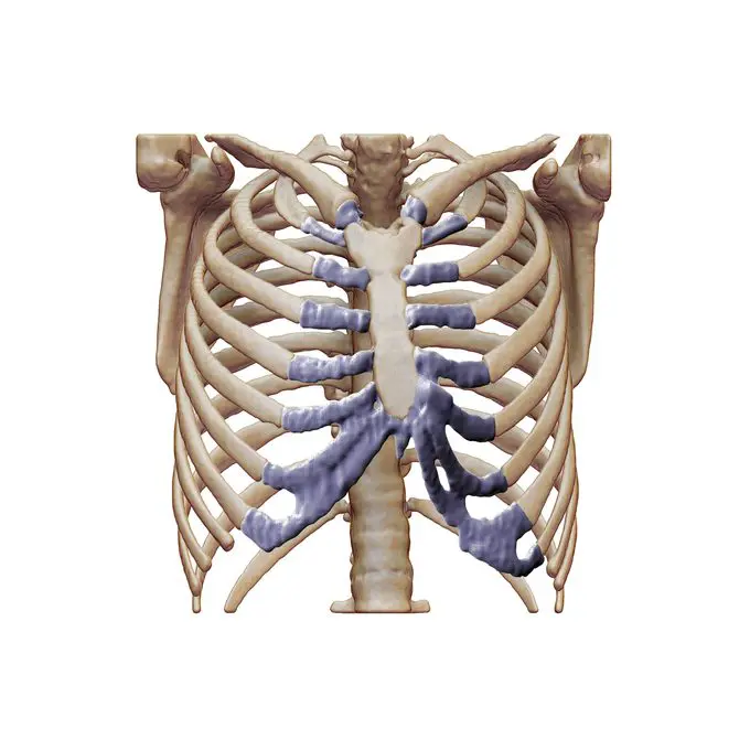
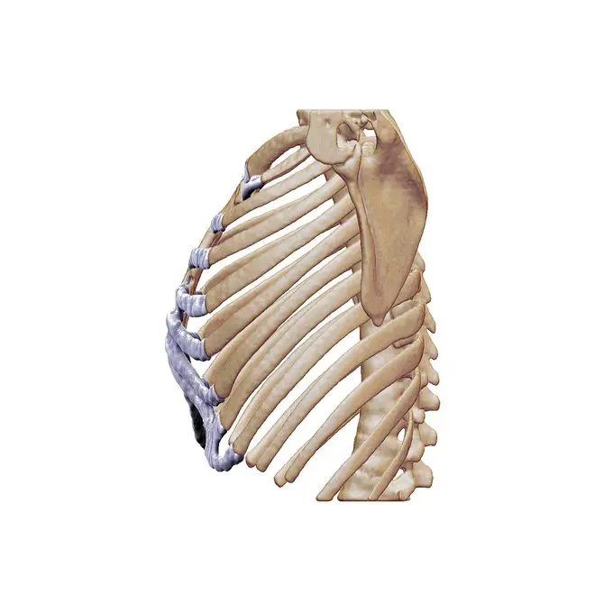
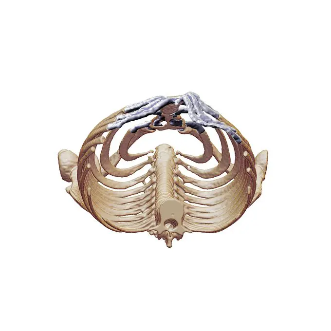
Pectus carinatum results from overgrowth of the costal cartilages — the flexible connective tissue joining the ribs to the sternum. Rather than growing in line with the chest wall, the cartilages elongate and curve anteriorly, displacing the sternum forward. The deformity is often asymmetric, with one side more prominent than the other. The CT reconstructions below demonstrate a left-sided asymmetric variant.
Physical symptoms
Many patients have no significant functional symptoms and present primarily due to concerns about appearance. When symptoms are present, they typically relate to the altered mechanics of the chest wall.
Visible chest prominence
The outward protrusion of the sternum and adjacent cartilages is the defining physical finding. It may be central and symmetrical, predominantly right-sided, or involve the upper sternum. Asymmetry is common and can be associated with sternal rotation.
Chest pain & tenderness
Aching or sharp chest pain is common, often worse when lying prone (face-down) or when pressure is applied to the sternum. Costochondral tenderness — pain at the junction of rib and cartilage — may be prominent.
Poor posture & spinal changes
Compensatory postural changes — rounded shoulders, thoracic kyphosis, and altered spinal curvature — frequently accompany pectus carinatum. Scoliosis is present in a proportion of patients and should be formally assessed.
Exertional breathlessness
Some patients describe reduced exercise tolerance or breathlessness during vigorous activity. The barrel-shaped chest wall can alter respiratory mechanics, increasing the effort required for breathing during exertion.
‘Asthma-like’ symptoms
Respiratory symptoms resembling asthma — wheeze, cough, or variable breathlessness — are described in up to a quarter of adolescents with pectus carinatum. These may be related to chest wall mechanics rather than true airway disease.
Palpitations
Some patients with pectus carinatum have an associated mitral valve prolapse, which can cause palpitations or awareness of the heartbeat. This warrants echocardiographic assessment if suspected, though it is often of no haemodynamic significance.
Psychological impact
The visible nature of pectus carinatum means psychological and social effects are frequently significant, particularly during adolescence. Many young patients report avoiding swimming, sport, or situations where their chest may be visible. Body image concerns, low self-esteem, and social withdrawal are common, and in a proportion of patients these reach clinically significant levels.
At the Pectus Clinic, psychological impact is assessed as part of the standard initial consultation. Treatment decisions take into account not only objective severity but also the individual’s experience of their condition. Psychological support is available within the multidisciplinary team where appropriate.
How severity and shape vary
Pectus carinatum is described by the area and shape of the chest wall prominence. The commonest form is chondrogladiolar, where the middle or lower sternum projects forward. A less common form is chondromanubrial, where the upper sternum is more prominent. The deformity may be symmetrical or asymmetrical, and mixed features of pectus carinatum and excavatum can coexist.
Associated findings are common. Rib flare — lateral flaring of the lower costal margin — frequently accompanies the central protrusion and can be a source of pain and self-consciousness in its own right. Postural changes, including thoracic kyphosis and forward shoulder posture, are seen in many patients and are addressed as part of a structured assessment.
The variation in presentation means that no single treatment approach fits all patients. Cartilage flexibility is a key determinant of treatment: in younger patients and adolescents, the cartilage remains pliable and bracing can achieve excellent correction; in skeletally mature patients, flexibility diminishes and surgical options become more relevant. Assessment is individualised, and treatment is planned accordingly.
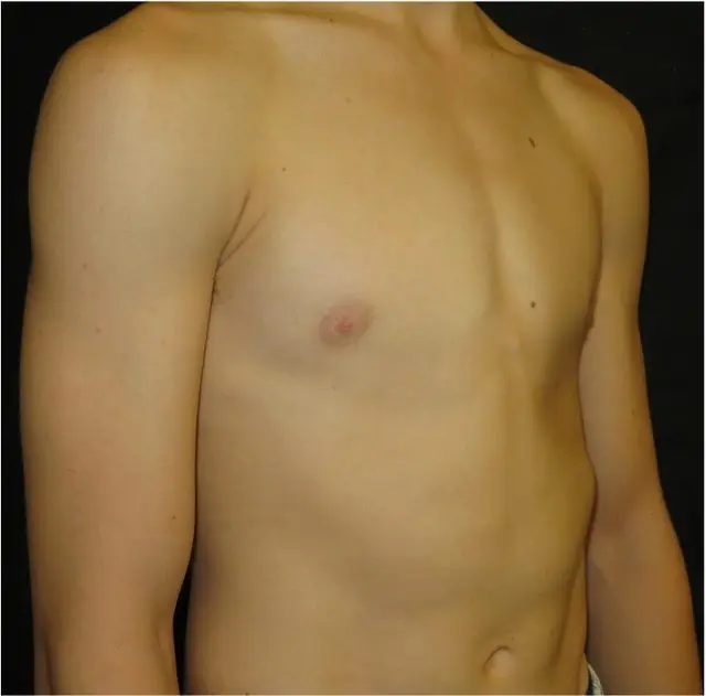
Mild symmetric
Low-grade central protrusion of the lower sternum with bilateral symmetry. Often first noticed by parents during early puberty. The cartilage is flexible at this stage, making bracing highly effective when started promptly.
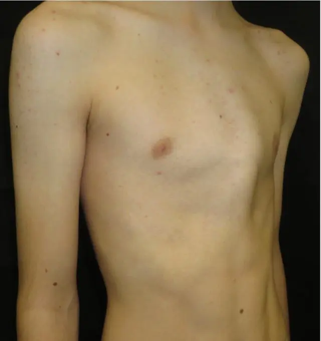
Asymmetric with sternal rotation
Unilateral protrusion with significant sternal rotation and contralateral flattening. If flexible, brace fitting must use a customised pad configuration to address the offset prominence.
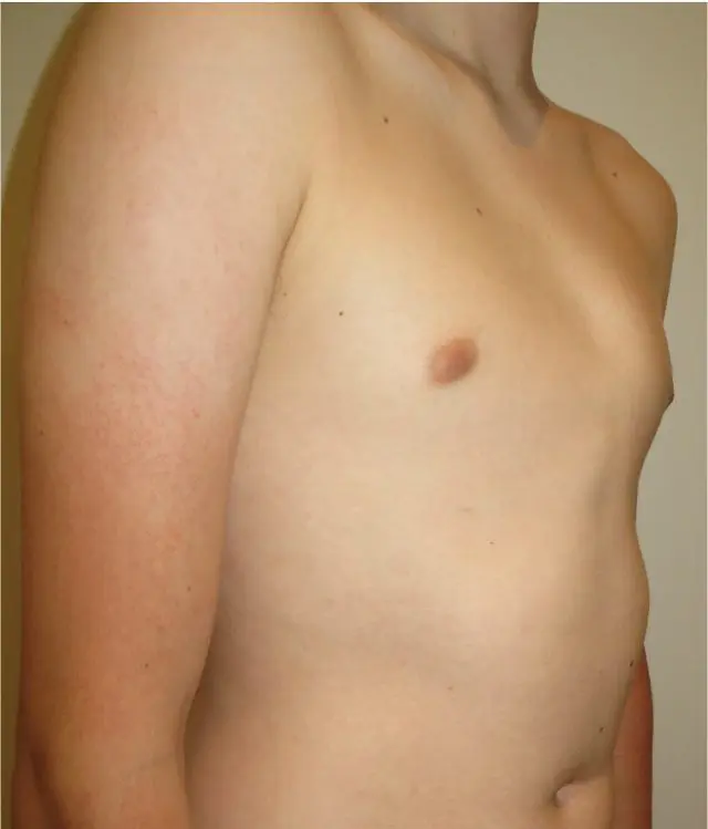
Severe symmetric (keel chest)
Pronounced central protrusion of the lower sternum and bilateral costal cartilages — the classic ‘keel chest’ pattern. In actively growing adolescents, full-time bracing achieves correction in over 90% of compliant patients.
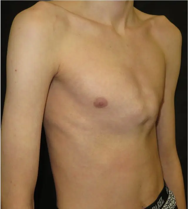
Extreme symmetric
Severe bilateral protrusion extending from the manubrium to the xiphisternum. In skeletally mature patients with this degree of deformity, surgical correction is often the most appropriate option. CT evaluation and multidisciplinary planning are essential.
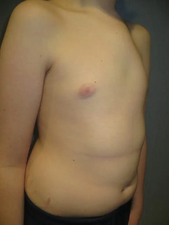
Superior (chondromanubrial) variant
A less common form in which the upper sternum and manubrium are most prominent, rather than the typical lower sternal region. Bracing is not usually possible due to the location of the prominence, which is too high for standard brace contact. CT evaluation is recommended before treatment planning, and surgical assessment is usually the appropriate next step if warranted.
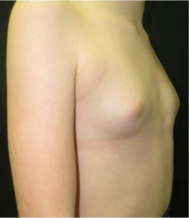
Female presentation
Pectus carinatum in female patients is less common (approximately 1 in 4 cases) but often presents later and with greater psychosocial impact. Brace design and surgical planning must account for breast tissue and the distinct chest wall anatomy in women.
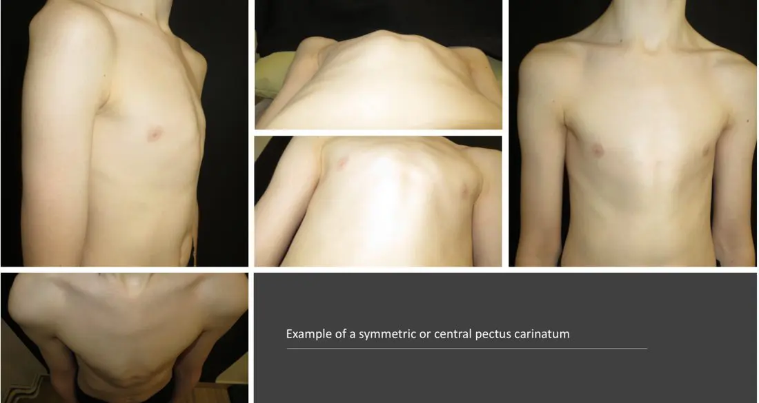
Symmetric (keel chest)
Equal protrusion on both sides with a central sternal peak. Often described as a ‘keel’ shape. Usually involves the lower sternum and bilateral costal cartilages. Responds well to bracing when growth is still occurring.
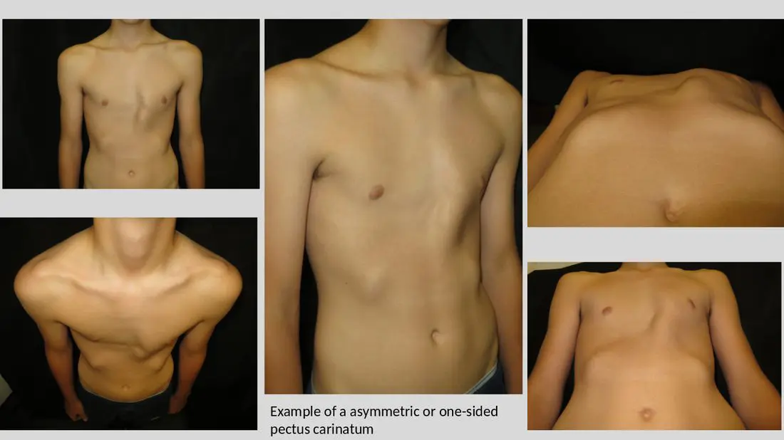
Asymmetric (lopsided)
The most common pattern. Protrusion is greater on one side — usually the right — often with associated sternal rotation and compensatory flattening or depression of the opposite side. May require asymmetric bracing or surgical planning.
How is pectus carinatum assessed?
Diagnosis is primarily clinical — careful physical examination by a specialist like Mr Hunt, who is familiar with chest wall deformity, is usually sufficient. Objective measurements and imaging are used to quantify severity, plan treatment, and monitor progress.
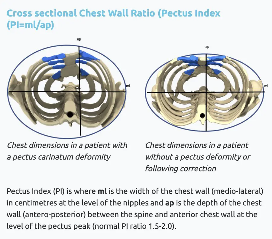
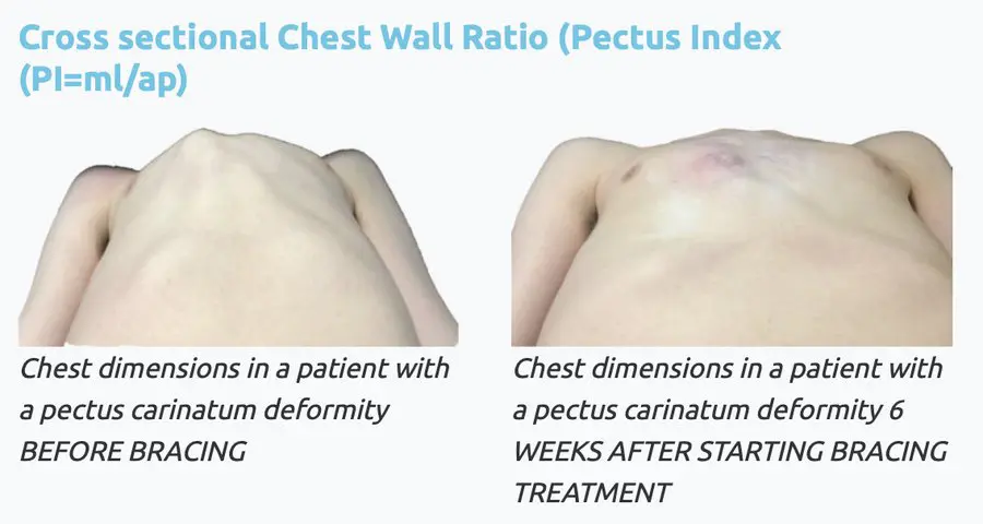
A PI nearer to 1.0 represents a more rounded, protruding shape indicating greater severity. Measurements are taken at the level of the nipples. Serial PI measurements track response to bracing.
Clinical assessment
Physical assessment by a chest wall specialist remains the cornerstone of diagnosis. The PI is measured using callipers at the nipple line. Serial measurements track treatment progress.
Clinical photography & 3D topographical scan
Standardised photographs (anterior, lateral, oblique views) are taken at initial assessment along with a 3D topographical scan and at each follow-up appointment, providing an objective visual record of change during bracing or after surgery.
CT chest (with 3D reconstruction)
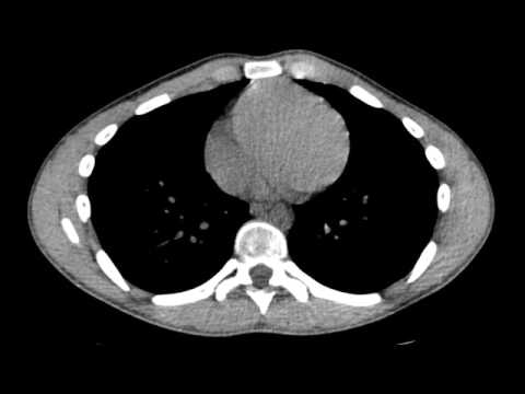
Provides detailed cross-sectional anatomy of the chest wall, quantifies sternal rotation, and is used in surgical planning. Not routinely required for straightforward bracing cases, but valuable in complex or asymmetric deformities and before corrective surgery.
Cardiac & pulmonary investigations
Where there are concerns about cardiac or respiratory symptoms — such as palpitations or breathlessness — further investigation may be appropriate. This can include echocardiography, formal lung function testing (spirometry), and cardiopulmonary exercise testing (CPET), guided by clinical assessment and symptom severity.
Conditions associated with pectus carinatum
A thorough assessment considers conditions that may coexist with the chest wall deformity. Identifying these influences treatment planning and informs the overall management approach.
Marfan syndrome
Pectus carinatum is a recognised skeletal feature of Marfan syndrome. Any patient with tall stature, long limbs, arachnodactyly, arm span exceeding height, or a family history of aortic dissection or sudden cardiac death should be assessed for Marfan syndrome, with cardiology and genetic review as appropriate.
Ehlers-Danlos & hypermobility
Generalised joint hypermobility and Ehlers-Danlos syndrome (particularly the hypermobile subtype) occur at increased rates in patients with chest wall deformities. This can affect posture, pain patterns, and response to physiotherapy. Awareness of underlying hypermobility is important when planning bracing and rehabilitation.
Poland syndrome
An uncommon condition involving hypoplasia of the pectoralis major and, in some cases, associated chest wall and rib abnormalities on one side. Pectus carinatum may occur in this context and is typically asymmetric, requiring tailored assessment and planning. Learn about Poland syndrome →
Scoliosis & spinal deformity
Lateral curvature of the spine occurs alongside pectus carinatum in a proportion of patients and should be assessed clinically and on plain radiographs. Significant scoliosis may influence brace design, treatment sequencing, and the approach to surgical correction.
Rib flare
Flaring of the lower costal margin is common in pectus carinatum, particularly with asymmetric deformities. It may persist after correction of the primary protrusion and should be assessed and addressed separately where it contributes to symptoms or appearance. Learn about rib flare →
Postural & musculoskeletal problems
Compensatory postural changes are common, including rounded shoulders, increased thoracic kyphosis, and pectoral muscle tightness. Physiotherapy and postural rehabilitation are an important part of the overall treatment pathway, both alongside bracing and following surgery.
Psychological well-being
Significant psychological impact — including anxiety, social avoidance, and body image disturbance — is common and well documented. These concerns often drive the decision to seek treatment. Psychological assessment and support are an integral part of the multidisciplinary approach at the Pectus Clinic.
Treating pectus carinatum
Treatment is tailored to each individual based on age, severity, skeletal maturity, cartilage flexibility, and the patient’s own goals. In growing children and adolescents, bracing is the preferred first-line approach. Surgery is reserved for those who are skeletally mature, have failed bracing, or have structural features not amenable to conservative correction.
External chest wall bracing
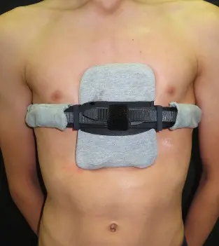
A custom-manufactured and fitted brace applies controlled anterior pressure to the sternal protrusion, gradually correcting the deformity by reshaping the flexible costal cartilages during active growth. Correction rates exceed 90% in well-selected, compliant adolescent patients.

Physiotherapy & postural rehabilitation
Targeted physiotherapy addresses the musculoskeletal consequences of pectus carinatum — including tight pectoral muscles, poor scapular control, thoracic kyphosis, and altered breathing mechanics. It is used alongside bracing and forms an important part of rehabilitation after surgery. A personalised programme is developed in collaboration with specialist physiotherapists.

Psychological support
For patients in whom the psychological impact of pectus carinatum is clinically significant, access to specialist psychological support is available within the multidisciplinary team. This may involve cognitive behavioural approaches, body image work, or support to address anxiety, avoidance, and school-related difficulties. Psychological wellbeing is assessed at every consultation.
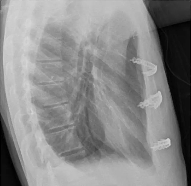
Modified Ravitch procedure
The modified Ravitch operation involves removal of the abnormally elongated costal cartilages through an anterior chest incision, along with cutting the sternum if necessary, allowing the chest to be repositioned. This is typically held in place with multiple titanium plates. The cartilages regenerate over several months. It is the most established surgical approach for pectus carinatum and can be tailored to address asymmetric or complex deformity patterns.
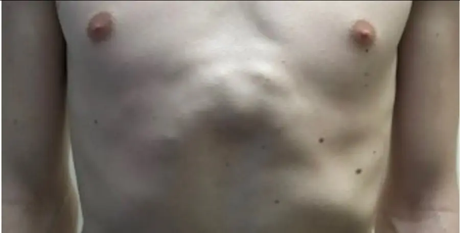
Mini-Ravitch procedure and Combined approach
In selected patients, a mini-Ravitch approach combines limited cartilage resection with or without plate-assisted sternal repositioning through smaller incisions. This reduces the operative footprint of open correction whilst addressing the structural deformity. Detailed CT planning and multidisciplinary review are essential for appropriate patient selection. As a combined approach, ongoing correction can be optimised using a bespoke external chest brace following surgery to correct the now more flexible chest deformity.
Verified patient recommendations
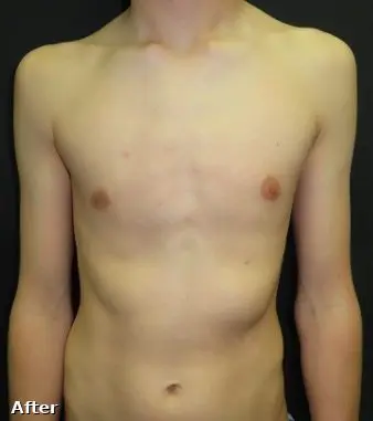
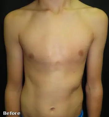
All patients shown have given consent for educational use. Results depend on age, deformity type, severity and treatment chosen.
“Wearing the brace has been a bit annoying but has still been 100% worth having!”
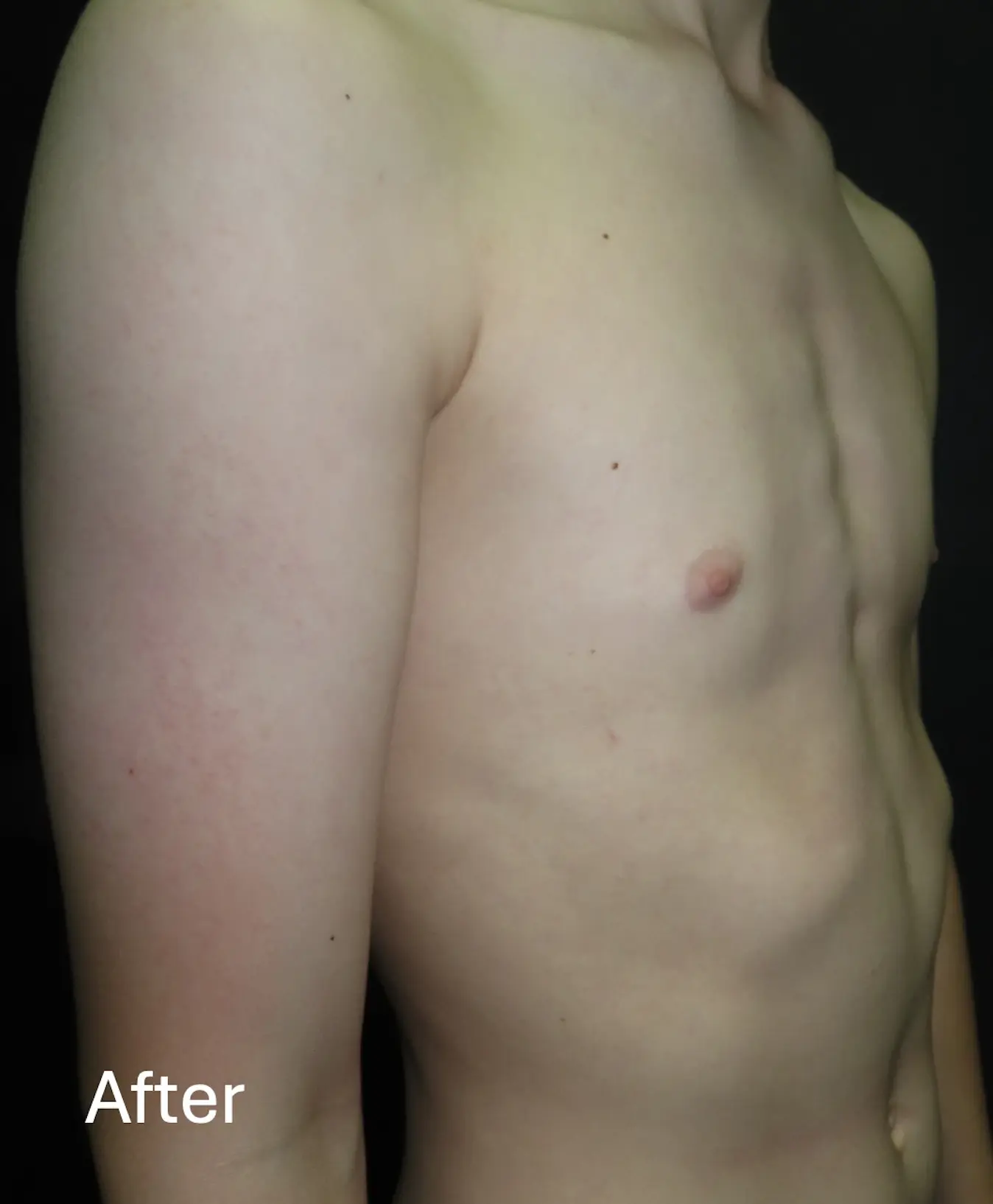
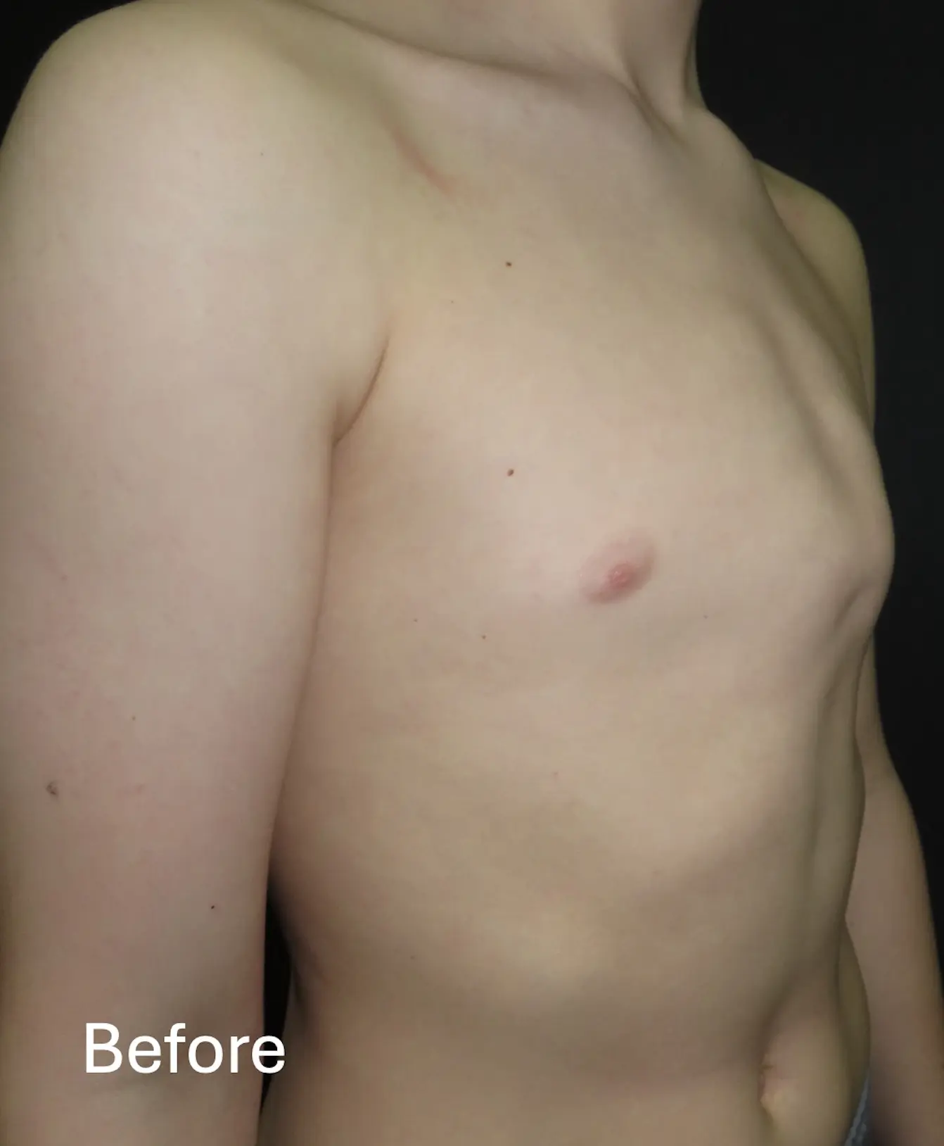
“This is a magical brace! Dr Hunt has been very patient and helpful, fully understanding my situation and prescribing the brace to perfectly remedy my chest. Thanks a ton!”
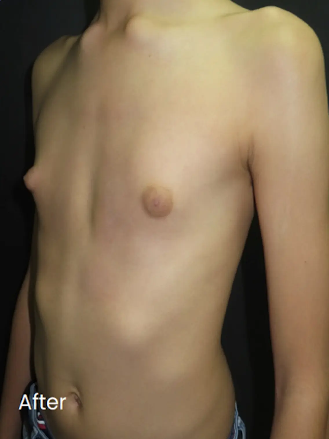
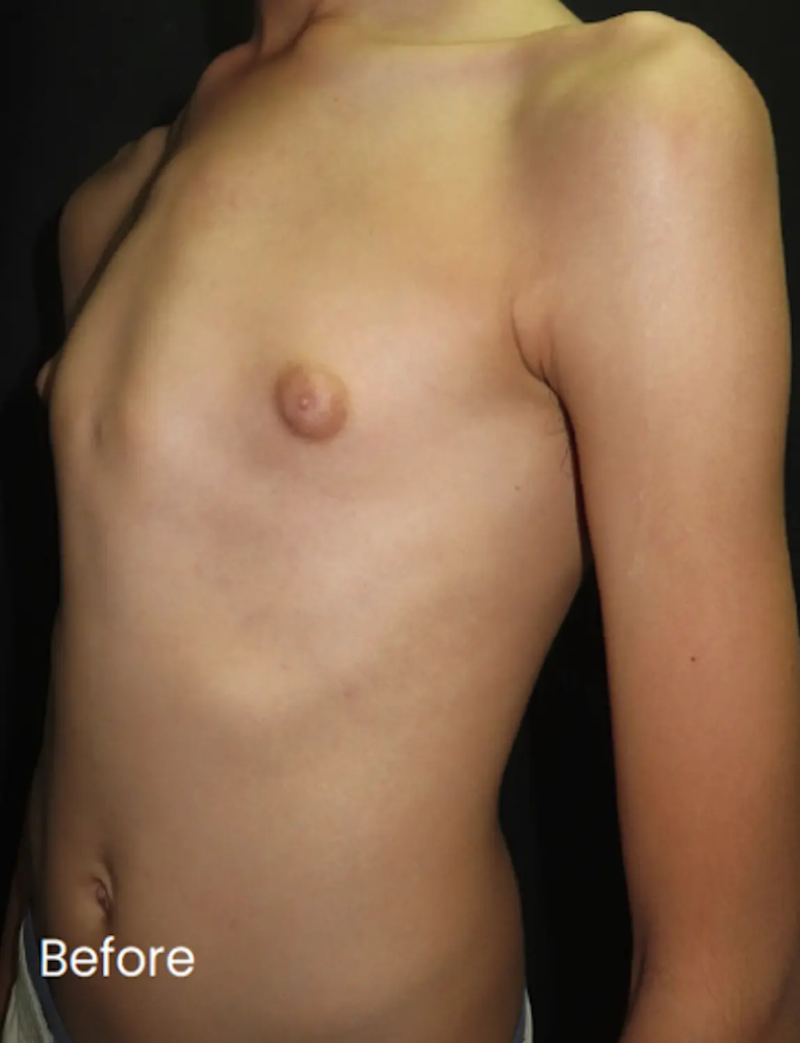
“Successfully corrected my chest.”
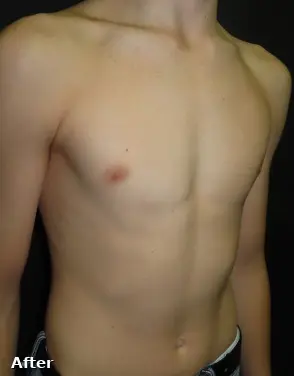
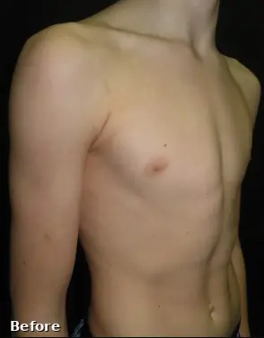
“I am happy with the overall results I have seen from the programme and have received loads of comments about ‘how much it has improved’.”
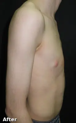
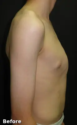
“Before bracing I thought surgery would be needed so I had no expectations, only worries about scarring. I’m so happy with the results and not having surgery — it has improved my mental wellbeing and overall confidence. Drastically.”
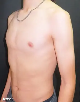
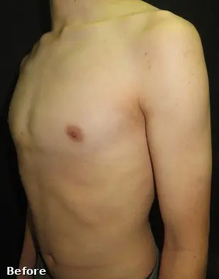
“Amazing, beyond any expectation I had before, would highly recommend to anyone with pectus. It's 100% worth it.”

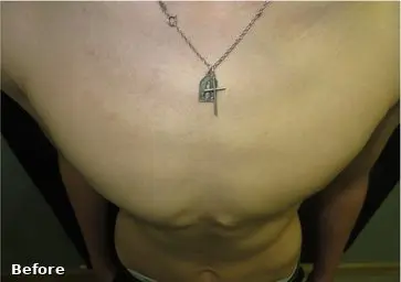
“This is the best thing I have done. It has honestly changed my life and I can't thank Dr Hunt enough.”
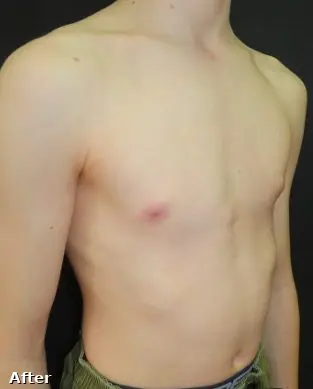
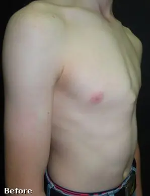
“Mr Hunt has encouraged me all the way through the programme. I have just returned from holiday and I was confident in taking my top off.”
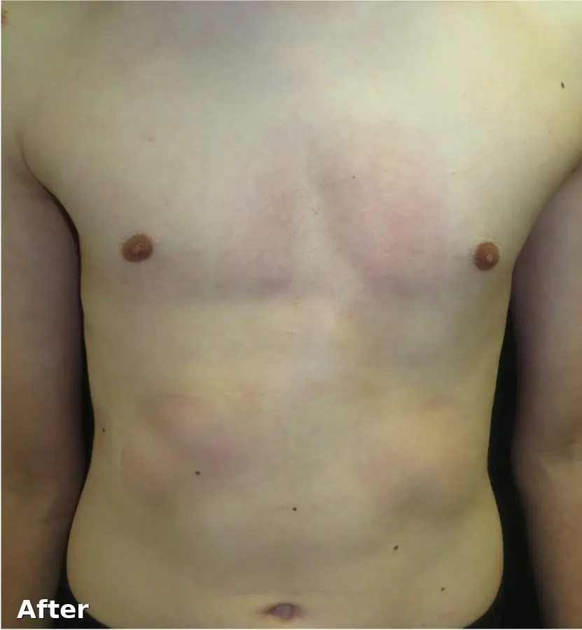
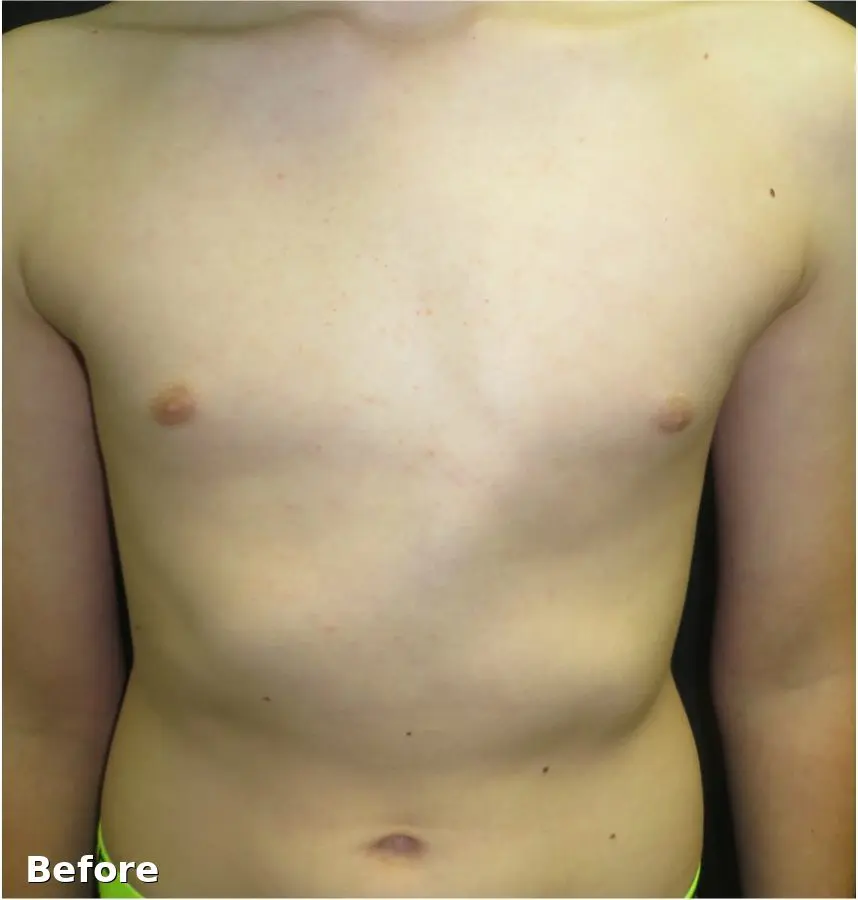
“The bracing experience is very easy and simple for an amazing outcome. The brace is comfortable and very well disguised under your shirt.”
Request an assessment
If you or your child has a chest wall protrusion that you would like assessed, we are happy to arrange an initial consultation. Assessment includes clinical examination, Pectus Index measurement, and a discussion of the most appropriate treatment pathway.